Understanding Melanoma: Why It Is Considered the Most Dangerous Type of Skin Cancer
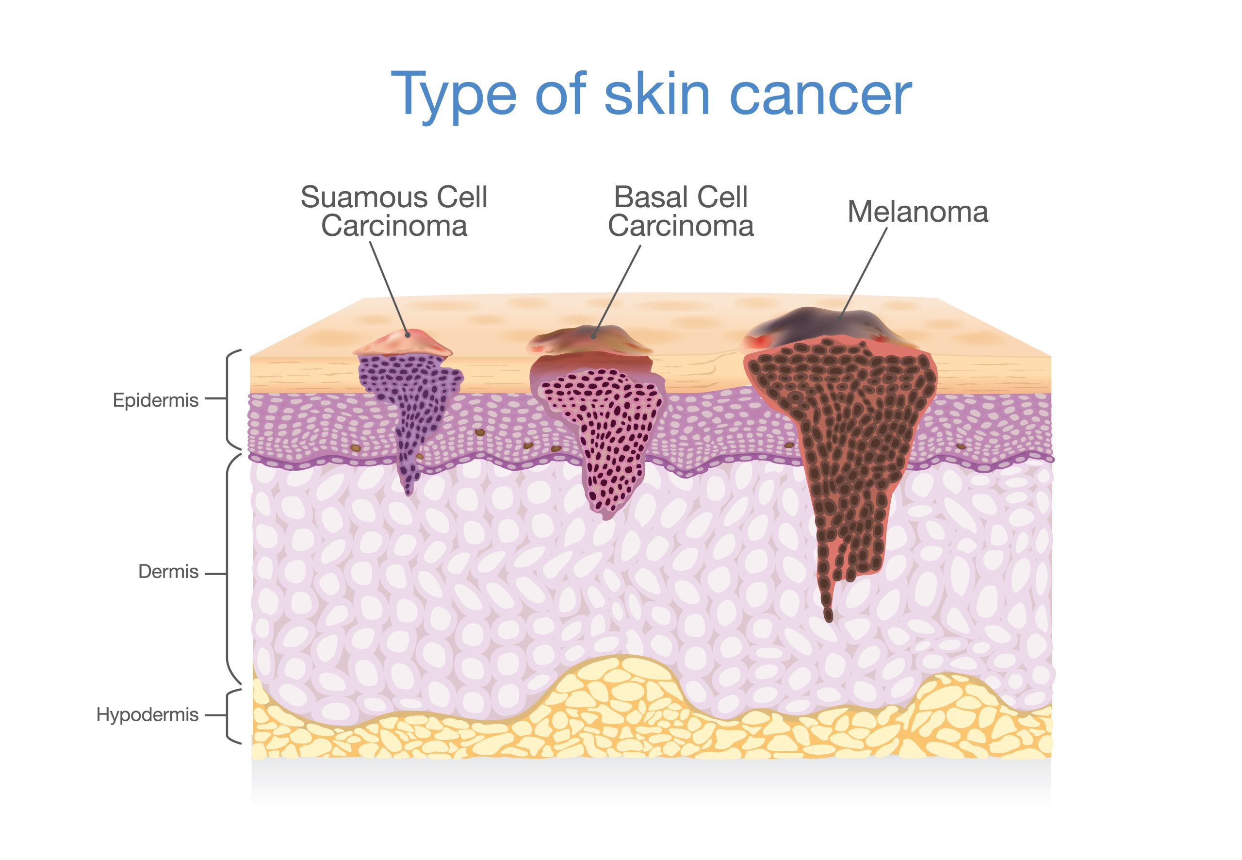
The most dangerous type of skin cancer is melanoma, a malignancy that develops in the melanocytes—the cells responsible for producing pigment in the skin. Unlike more common non-melanoma skin cancers, melanoma is highly aggressive because of its significant propensity to metastasize, or spread, to distant organs, including the brain, lungs, and liver, if it is not detected and treated during its early stages.
The Biological Basis of Melanoma's Lethality
To understand why melanoma occupies the top position in terms of danger among cutaneous malignancies, one must look at the specific biology of melanocytes. These cells are located in the basal layer of the epidermis and are derived from the neural crest, an embryonic tissue that migrates widely throughout the body during development. Because of this migratory heritage, melanocytes maintain a biological plasticity that can be exploited by cancerous mutations.
When these cells transform into malignant melanoma, they often retain the ability to invade surrounding tissues and navigate the lymphatic and circulatory systems with relative ease. While basal cell carcinoma and squamous cell carcinoma—the two most common forms of skin cancer—tend to remain localized or grow slowly, melanoma can penetrate deeper into the dermis and reach the subcutaneous fat, where it gains access to the dense network of blood vessels and lymph nodes. Once the cancer enters the bloodstream, it can seed tumors in virtually any organ system, making systemic management significantly more complex than the localized surgical excision often used for other skin cancers.
Differentiating Skin Cancers: A Comparative Overview
Medical literature classifies skin cancers into two broad categories: non-melanoma skin cancer (NMSC) and melanoma. Understanding this distinction is essential for recognizing why the medical community emphasizes the urgency of identifying melanoma.
| Feature | Basal Cell Carcinoma (BCC) | Squamous Cell Carcinoma (SCC) | Melanoma |
|---|---|---|---|
| Origin Cell | Basal cells | Squamous cells | Melanocytes |
| Metastasis Risk | Very Low | Low (but possible) | High |
| Growth Rate | Slow | Moderate | Fast/Variable |
| Common Location | Sun-exposed areas | Sun-exposed areas | Anywhere on the body |
| Primary Danger | Local tissue destruction | Local invasion | Systemic metastasis |
As illustrated in the table, the primary differentiator is the metastatic potential. While basal cell and squamous cell carcinomas are far more frequent in the general population, their mortality rates remain low because they are typically caught before they invade underlying structures. Melanoma, conversely, can progress from an in-situ lesion to a life-threatening, systemic disease in a relatively short window of time.
Risk Factors and Preventative Awareness
The development of melanoma is multifactorial, involving a combination of genetic predisposition and environmental triggers. As of 2026, clinical guidelines continue to stress that intermittent, intense exposure to ultraviolet (UV) radiation—often resulting in blistering sunburns, particularly during childhood or adolescence—is one of the most significant environmental risk factors.
Beyond UV exposure, individuals with a high number of atypical moles (dysplastic nevi) or a personal or family history of melanoma are at an elevated risk. Other factors include having a fair complexion, light eye color, or a weakened immune system. It is important to note that while UV radiation is a major culprit, melanoma can also occur in areas of the body that receive little to no sun exposure, such as the palms of the hands, the soles of the feet, or under the fingernails. This phenomenon, known as acral lentiginous melanoma, is particularly common in individuals with darker skin tones, suggesting that skin cancer awareness should be a universal health priority rather than one limited to specific demographics.
The Importance of the ABCDE Rule
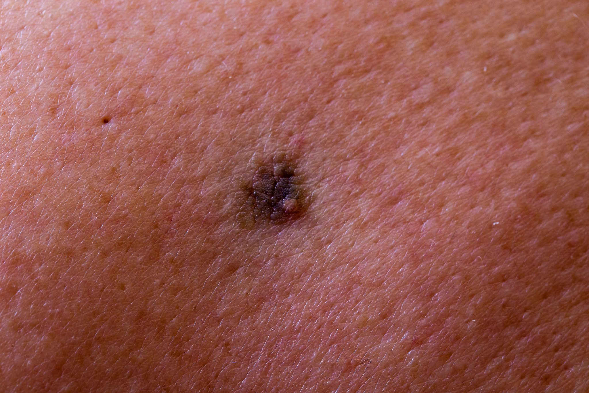
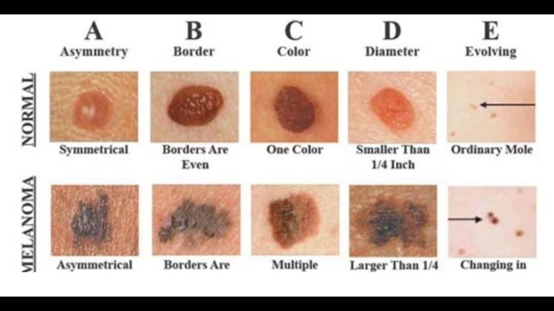
Early detection remains the single most effective tool in reducing mortality from melanoma. Dermatologists utilize a standardized screening method known as the ABCDE rule to help individuals distinguish between benign moles and potentially dangerous lesions:
- Asymmetry: If one half of the mole does not match the other half, it warrants professional examination.
- Border: Benign moles usually have smooth, distinct edges. Melanoma borders are often ragged, notched, or blurred.
- Color: A single, uniform color is typically a positive sign. A lesion that displays multiple shades—including brown, black, tan, red, or blue—is a significant warning sign.
- Diameter: While not an absolute rule, a lesion larger than the eraser of a standard pencil (approximately 6 millimeters) is often flagged for closer inspection.
- Evolving: Any change in size, shape, elevation, or color of an existing mole, or the development of new symptoms such as bleeding, itching, or crusting, requires immediate medical consultation.
Diagnostic Procedures and Staging
When a healthcare provider suspects a lesion is malignant, the standard diagnostic procedure is a skin biopsy. This is a minor surgical process where a portion or the entire lesion is removed and sent to a pathologist. The pathologist examines the tissue under a microscope to determine if the lesion is malignant and, if so, how deep it has grown into the skin (Breslow thickness).
Staging follows the biopsy and is crucial for determining the treatment plan. Staging in 2026 utilizes the AJCC (American Joint Committee on Cancer) system, which evaluates the tumor's thickness, whether it has ulcerated, whether it has spread to nearby lymph nodes, and whether it has metastasized to distant organs. Early-stage (Stage I or II) melanoma is primarily treated via wide excision, where the tumor and a margin of healthy skin are removed. Higher stages often involve multidisciplinary care, including specialized imaging (such as PET or CT scans), sentinel lymph node biopsy, immunotherapy, targeted therapy, or, in specific cases, surgical intervention for metastatic sites.
Advancements in Treatment and Prognosis
The prognosis for melanoma has improved significantly over the last decade due to the introduction of systemic therapies. Immunotherapy agents, such as checkpoint inhibitors, work by "taking the brakes off" the immune system, allowing it to recognize and attack melanoma cells that were previously "hidden" from detection. Similarly, targeted therapies can be used in patients whose tumors harbor specific genetic mutations, such as the BRAF mutation, effectively blocking the proteins that drive the cancer’s growth.
Despite these advancements, the effectiveness of these treatments remains highly dependent on the stage of the disease at the time of diagnosis. While metastatic melanoma was once considered largely untreatable, current options offer significantly extended survival and quality of life for many patients. Nevertheless, the physical, emotional, and financial burdens of treating advanced skin cancer underscore why the "most dangerous" label is not just a statistical classification, but a call to action for proactive skin health.
Frequently Asked Questions
Is melanoma the only type of skin cancer I should worry about? No. While melanoma is the most dangerous, other forms like squamous cell carcinoma can also be aggressive if left untreated. Regular skin checks should screen for all changes in the skin, not just those that look like melanoma.
Can sunscreen prevent all types of skin cancer? Sunscreen is a vital component of sun protection, but it is not 100% effective. It should be used alongside other behaviors, such as seeking shade, wearing protective clothing, and avoiding sun exposure during peak UV hours (typically 10 a.m. to 4 p.m.).
Do I need a professional skin exam if I don't have many moles? Yes. While high-risk individuals should have more frequent exams, the American Academy of Dermatology recommends that everyone be aware of their skin. Even those with few moles can develop skin cancer, as it can appear in previously clear skin or areas rarely exposed to the sun.
What should I do if I notice a change in a mole? Do not wait for it to change further. Schedule an appointment with a board-certified dermatologist for a clinical examination. Early detection is the most important factor in the success of skin cancer treatment.
Is tanning bed use linked to melanoma? Yes. The use of indoor tanning beds is strongly linked to an increased risk of developing melanoma. The intense UV radiation delivered by these devices causes cumulative DNA damage in skin cells, and many medical organizations strongly advise against their use.
Recommendations for Next Actions
If you are concerned about a specific lesion, the most important step is to document its appearance and schedule a consultation with a board-certified dermatologist. Do not attempt to treat or remove a suspicious lesion yourself, as this can delay accurate diagnosis and potentially complicate future pathological assessment. For individuals with a family history of melanoma or a high personal history of sun damage, establish a baseline "total body skin examination" with a medical professional. This session will provide a professional record of your skin’s current state, making it easier to identify future changes. Practicing monthly self-exams using the ABCDE guidelines will allow you to act quickly if you notice anything unusual.
Image Gallery
Select an image to open it in a new tab.



Declan O'Connor
Higher Education and Campus Culture Reporter
