Understanding Braces and MRI Safety: Clinical Guidelines and Patient Precautions
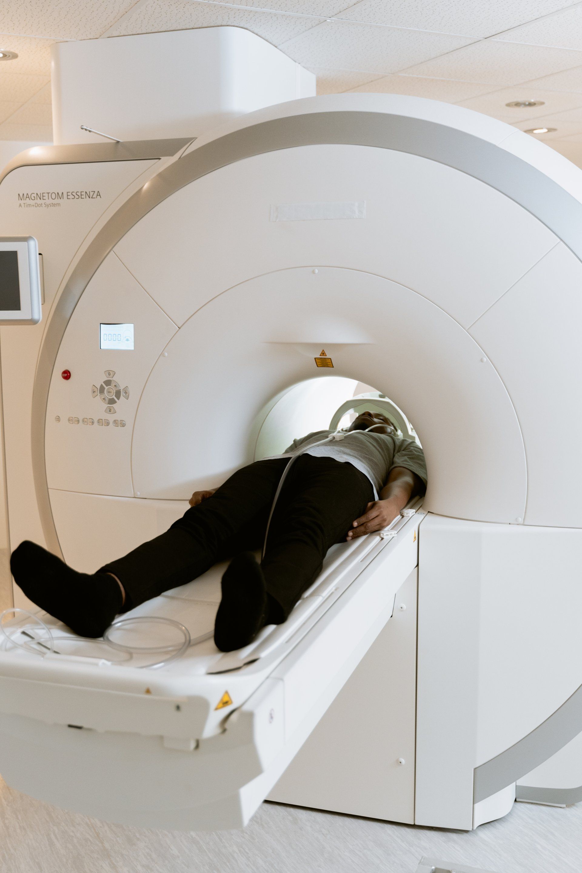
Magnetic Resonance Imaging (MRI) is a vital diagnostic tool, but its reliance on powerful magnetic fields necessitates rigorous safety protocols, especially for patients with metallic hardware. Orthodontic braces, which often contain stainless steel, nickel-titanium, or cobalt-chromium alloys, present unique challenges in an MRI environment. While most patients with braces can safely undergo an MRI, the primary risks involve image distortion artifacts, potential heating of the metal components, and—in extremely rare cases—the mechanical interaction between the scanner’s magnetic field and the orthodontic appliances.
The Physics of MRI and Metallic Interference
An MRI machine operates by creating an extremely strong, constant static magnetic field, typically measured in Teslas (T). Common clinical scanners operate at 1.5T or 3.0T. When an object containing ferromagnetic or paramagnetic materials enters this field, several physical phenomena occur that impact diagnostic quality and patient safety.
First, metallic materials, including the stainless steel used in many orthodontic brackets, can cause localized distortion of the magnetic field. Because MRI relies on the homogeneity of the magnetic field to create images, the presence of metal induces a "susceptibility artifact." This appears on the scan as a "signal void" or "blooming artifact," where the area around the metal appears black, distorted, or significantly blurred. In the context of head and neck imaging, this can obscure critical anatomy, such as the brain, orbits, or salivary glands, making a diagnosis difficult or impossible.
Second, the interaction between the radiofrequency (RF) pulses used in MRI and the metal in braces can lead to induced currents. In theory, this could result in localized heating of the orthodontic wires or brackets. However, because orthodontic appliances are usually small and integrated into the soft tissues of the mouth, the risk of clinically significant thermal injury is exceptionally low, provided the patient does not experience sudden, sharp sensations of heat.
Assessing the Risk: Ferromagnetic vs. Non-Ferromagnetic Materials
Not all metal used in orthodontics reacts the same way. The level of danger is determined by the material composition of the braces.
- Stainless Steel: Widely used for brackets and archwires, stainless steel is generally paramagnetic or weakly ferromagnetic. While it may cause significant image artifacts, it typically does not exert enough mechanical force in a 1.5T or 3.0T field to cause the brackets to detach or move.
- Nickel-Titanium (NiTi): These wires are used for their "shape memory" properties. They are generally considered safe regarding magnetic displacement, though they can still produce image artifacts.
- Ceramic and Composite Brackets: These are non-metallic and theoretically cause fewer artifacts. However, it is vital to note that even ceramic brackets often have a metal slot or a metallic base, and the archwire running through them is almost always metallic. Therefore, "non-metallic" braces are rarely completely free of MRI-related interference.
Clinical Protocols and The Importance of Communication
Before undergoing an MRI, it is standard practice to complete a detailed safety screening form. You must disclose the presence of braces to the MRI technologist and the radiologist.
The decision to proceed with an MRI while wearing braces is ultimately made by the radiologist, who weighs the diagnostic necessity of the scan against the anticipated severity of the artifacts. In many cases, if the area of interest is the brain or the cervical spine, the artifacts from the braces may be so severe that they render the scan inconclusive. If the target area is in the abdomen or lower extremities, the braces might be considered far enough away from the "isocenter" (the center of the magnet) that they cause minimal disruption to the image quality.
Steps for Patients Preparing for an MRI with Braces
- Advance Notification: Inform your healthcare provider and the MRI facility about your braces at least 48 hours before your scheduled appointment.
- Consult Your Orthodontist: If you are unsure of the material composition of your appliances, contact your orthodontist. They can provide documentation regarding the materials used, which the radiology department may keep on file.
- The Pre-Scan Screening: Be prepared for the technologist to perform a physical assessment of the mouth. They may ask you to remove any removable orthodontic appliances, such as retainers or clear aligners, which should be taken out regardless of their composition to avoid damage or loss.
- During the Scan: If you experience any unusual sensation—such as pulling, heating, or pain near your teeth—alert the technologist immediately using the provided call button.
Comparative Impact of Braces on MRI Quality
The following table summarizes the typical considerations for patients with orthodontic hardware undergoing an MRI.
| Appliance Type | Risk of Displacement | Image Artifact Severity | Diagnostic Impact |
|---|---|---|---|
| Stainless Steel Braces | Very Low | High | Severe in head/neck scans |
| Nickel-Titanium Wires | Negligible | Moderate | Minimal to moderate |
| Ceramic Brackets (w/ Metal Slot) | Very Low | Moderate | Moderate in head/neck scans |
| Removable Retainers (Plastic) | None | None | None (must be removed) |
| Lingual Braces (Behind teeth) | Very Low | High | Significant in head/neck scans |
Troubleshooting: What if the Scan is Obstructed?
If you arrive for your appointment and the radiologist determines that your braces will cause excessive artifacts, they may suggest alternatives. One option is to delay the MRI until the braces are removed. However, for most patients, this is not a practical solution.
If the diagnostic task is urgent and the braces are the primary source of interference, the radiologist may attempt to adjust the "sequence parameters" of the MRI. By changing the way the machine collects data—specifically by using "spin-echo" sequences instead of "gradient-echo" sequences—they can sometimes reduce the visual footprint of metallic artifacts. These adjustments require a high level of technical expertise and may increase the duration of the scan.
In cases where the artifacts are unavoidable and the diagnosis remains unclear, your physician may recommend an alternative imaging modality, such as a CT (Computed Tomography) scan. While CT scans involve low doses of radiation, they are not sensitive to the magnetic properties of metal and provide excellent imaging of bony structures, even when metallic hardware is present.
Frequently Asked Questions
Does the MRI pull the braces off my teeth? No. While there is a magnetic force, the bond strength of modern orthodontic adhesives is significantly higher than the force exerted by the magnet on the small mass of a metal bracket. There are no recorded instances of standard orthodontic brackets being "ripped" off by an MRI machine.
Do I need to remove my clear aligners or plastic retainers? Yes. You should always remove all removable dental appliances before entering the MRI room. While they are non-metallic and safe, they can be lost or damaged during the procedure, and they may still cause minor interference with the localized magnetic field.
Will my braces heat up during the scan? Some patients report a mild tingling or a sensation of warmth. This is due to the radiofrequency pulses. If this occurs, it is usually harmless, but if the sensation becomes uncomfortable, you must signal the technologist immediately to pause the scan.
Can I get an MRI if I have a permanent bonded retainer? Yes. Permanent (fixed) lingual retainers are usually made of thin stainless steel or braided wire. They are very small and typically cause minimal artifacts, especially since they are located far from the brain tissue in most imaging scenarios.
Is 3.0T MRI more dangerous for braces than 1.5T MRI? Higher field strengths, such as 3.0T, produce stronger artifacts because the susceptibility effects scale with the magnetic field strength. While the safety risk regarding physical displacement remains low, the image quality is often worse at 3.0T compared to 1.5T when metal hardware is present.
Practical Recommendations for Patients
If you are scheduled for an MRI, your best course of action is transparency. Do not attempt to hide the fact that you have braces, even if you believe they are "small" or "not that metallic." Radiology departments have established safety protocols specifically designed to handle metallic hardware. By providing accurate information about your orthodontic treatment, you allow the radiologist to tailor the scan protocols to your specific needs, potentially avoiding the need for a repeat scan.
Always carry your current medical records and, if possible, information from your orthodontist regarding the metal content of your appliances. If you have been told that your upcoming MRI is of the head or neck area, be mentally prepared for the possibility that the images might show some distortion. Open communication with the radiology team during the screening process is the most effective way to ensure both your safety and the diagnostic accuracy of your medical procedure.

Image Gallery
Select an image to open it in a new tab.




Akira Tanaka
International Academic Trends Correspondent
