
Microinvasive squamous cell carcinoma (MISCC) represents a clinical threshold in oncology where precancerous changes—specifically squamous cell carcinoma in situ—begin to penetrate the basement membrane of the epithelium. This localized invasion, typically defined by a depth of penetration into the underlying stroma of 3 millimeters or less, marks the transition from non-invasive disease to potentially metastatic cancer. Because it exists at the earliest stage of invasion, MISCC requires precise diagnostic evaluation to determine the most effective surgical or topical treatment pathway, balancing the need for complete clearance with the desire to minimize structural or functional damage to the affected tissue.
The Biological Definition of Microinvasion
To understand microinvasive squamous cell carcinoma, one must first distinguish between "in situ" disease and invasive disease. Squamous cells are the flat, scale-like cells that line various surfaces of the body, including the skin, the cervix, the oral cavity, and the esophageal lining. In squamous cell carcinoma in situ, abnormal cells are confined strictly to the epidermis or the surface layer; the basement membrane, which acts as a structural barrier between the surface and the deeper tissues (the stroma), remains intact.
Microinvasion occurs when these malignant cells breach this basement membrane. In a clinical and pathological sense, "micro" refers to the depth of this breach. While definitions can vary slightly depending on the anatomical site, most medical guidelines define microinvasion as an invasive component extending no more than 3 millimeters beyond the basement membrane. This specific measurement is critical because, at this depth, the risk of the cancer spreading to local lymph nodes or distant organs remains statistically very low. However, the presence of any invasion at all categorizes the lesion as a cancer rather than a high-grade dysplasia, fundamentally altering the clinical management strategy.
Anatomical Context and Site-Specific Variations
While the microscopic pathology of microinvasive squamous cell carcinoma is similar regardless of location, the clinical implications vary significantly based on where the lesion appears.
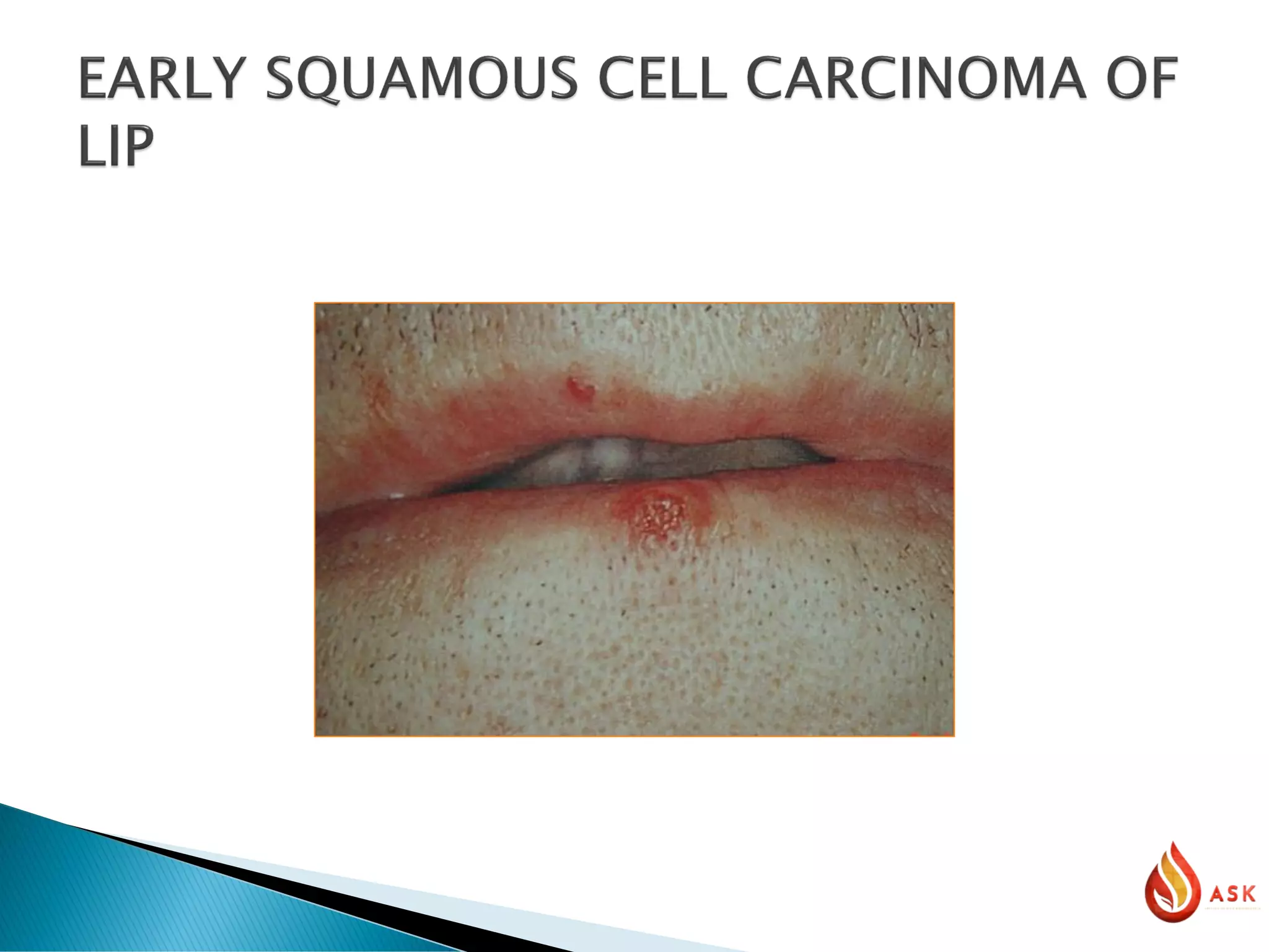
- Dermatological Context: In the skin, microinvasive squamous cell carcinoma is often a result of long-term ultraviolet (UV) exposure. It is frequently associated with actinic keratosis—a rough, scaly patch on the skin caused by years of sun damage. Here, the primary risk is local extension; cutaneous MISCC is generally highly curable with minor surgical intervention.
- Gynecological Context: Cervical microinvasive squamous cell carcinoma is a distinct staging category under the International Federation of Gynecology and Obstetrics (FIGO) guidelines. Because the cervix is a high-risk area for lymphatic spread, the assessment of depth is extremely rigorous. Even at a microinvasive level, the detection of lymphovascular space invasion (LVSI)—where cancer cells are found inside tiny blood or lymphatic vessels—can escalate the diagnosis from a simple localized lesion to one requiring more aggressive surgery.
- Head and Neck/Oral Context: In the mouth or throat, MISCC is highly sensitive to the patient’s history of tobacco and alcohol use. Because of the density of blood vessels and nerves in these areas, even "micro" invasion can have functional consequences on speech or swallowing if not treated with precision.
Diagnostic Verification and Pathological Staging
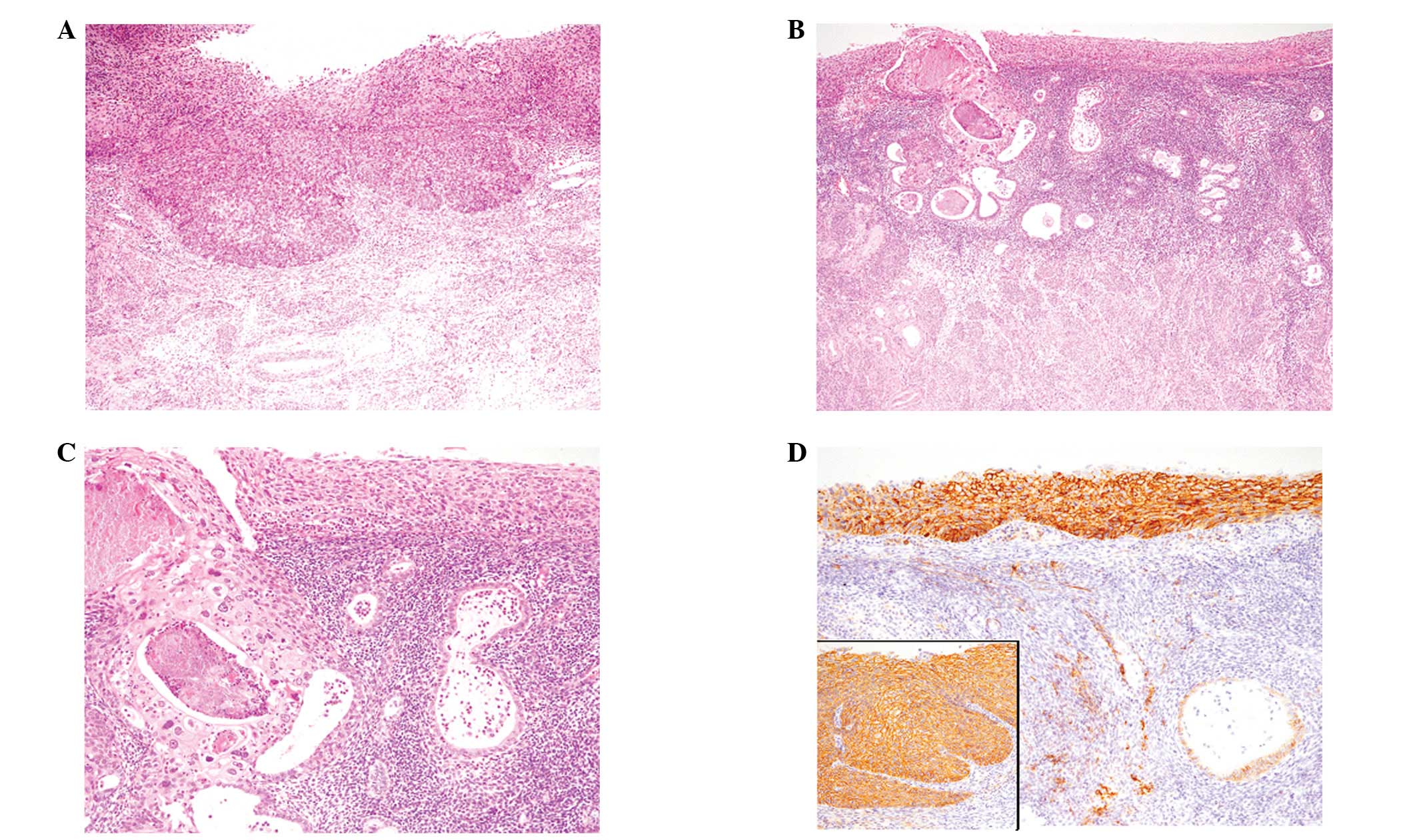
The diagnosis of MISCC cannot be confirmed through visual inspection alone; it requires a biopsy. A pathologist must examine the tissue sample under a microscope to confirm that the malignant cells have indeed breached the basement membrane.
The diagnostic process involves several key steps:
- Excision or Punch Biopsy: The clinician removes all or part of the suspicious lesion.
- Histopathological Analysis: The tissue is processed, sectioned, and stained (usually with hematoxylin and eosin). The pathologist measures the vertical depth of the invasion from the surface or the base of the epithelial crypt.
- Margin Assessment: The pathologist identifies whether the "margins" of the biopsy sample are "clear" or "positive." Clear margins mean that the surrounding tissue is free of cancer cells, suggesting that the entire lesion was removed. Positive margins suggest that the cancer may still be present at the edges of the original site, necessitating further treatment.
- LVSI Screening: The pathologist specifically checks for cancer cells within small vascular channels, as this is a high-risk indicator even when the depth of invasion is shallow.
Comparative Management Strategies
The management of MISCC is guided by the goal of achieving "negative margins" while preserving the maximum amount of healthy tissue. The following table summarizes standard approaches based on site and clinical necessity.
| Feature | Cutaneous (Skin) MISCC | Cervical MISCC | Oral/Mucosal MISCC |
|---|---|---|---|
| Primary Goal | Complete excision with margin control | Conservation of organ function | Elimination of risk factors & removal |
| Common Modality | Mohs Micrographic Surgery | Cone biopsy or LEEP | Wide local excision |
| Recurrence Risk | Low (if margins are clear) | Moderate (requires monitoring) | Higher (linked to habit cessation) |
| Primary Follow-up | Dermatological skin checks | Colposcopy/Pap smears | Clinical/Imaging evaluation |
Surgical and Non-Surgical Treatment Pathways
Treatment selection is based on the patient’s overall health, the exact depth of the invasion, and the potential for functional morbidity.
Surgical Excision
In most cases, surgical excision remains the gold standard. For skin lesions, Mohs micrographic surgery is frequently utilized. This technique involves removing the tissue in thin layers and examining each layer under a microscope while the patient waits. This ensures that every bit of cancer is removed while sparing as much healthy surrounding tissue as possible. This is particularly vital on the face, ears, or hands.
Topical Therapies
For very specific types of early-stage cutaneous MISCC, topicals such as Imiquimod or 5-fluorouracil may be considered in patients who are not candidates for surgery due to health complications. However, these are rarely the first-line choice because they do not allow for the histological confirmation of margins that surgery provides.
Specialized Procedures
In cervical cases, the Loop Electrosurgical Excision Procedure (LEEP) or cold-knife conization allows for the removal of the cervical transition zone. These procedures are diagnostic as well as therapeutic, as they provide the pathologist with a large enough tissue sample to definitively stage the microinvasion.
The Role of Lymphovascular Space Invasion (LVSI)
One of the most critical factors that can complicate a case of microinvasive squamous cell carcinoma is the presence of lymphovascular space invasion (LVSI). Even if the invasion is measured at less than 1 millimeter, the presence of tumor cells within a vessel significantly increases the probability that the cancer has already begun to migrate to the regional lymph nodes.
When LVSI is noted in a pathology report, the clinical management changes immediately. The lesion is no longer viewed simply as a localized "micro" issue. Oncologists may shift from a strategy of observation to one of wider surgical margins, potential sentinel lymph node biopsy, or closer imaging follow-up to detect potential recurrences early. Patients should ensure they receive a full copy of their pathology report and discuss the presence or absence of LVSI directly with their surgeon or oncologist.
Navigating the Psychological and Practical Impact
A diagnosis of "microinvasive" cancer often leaves patients in a state of confusion. They may hear the word "carcinoma" (cancer) but are told it is "microinvasive" (early stage), leading to conflicting feelings about the severity of the situation.
It is important to understand that while MISCC is serious, it is a highly treatable form of cancer. Because the diagnosis is made early, the prognosis is usually excellent. However, the primary practical challenge is long-term compliance with follow-up protocols. Patients with MISCC, especially those involving the skin or cervix, often have underlying environmental or viral triggers (such as high-risk HPV for cervical lesions or cumulative UV damage for skin lesions). Treatment does not resolve these underlying systemic risks; therefore, consistent check-ups are the most critical component of the "post-treatment" phase.
Common Questions and Expert Guidance
1. Is microinvasive squamous cell carcinoma the same as stage 1 cancer? Not necessarily. While it is an invasive cancer, clinical staging systems (like the TNM system) use specific criteria for "Stage 1." MISCC is a descriptive, pathological term that helps determine where that cancer falls within a staging system. It is usually considered the earliest possible stage of invasive cancer.
2. Can I wait a few months to treat my MISCC? Generally, no. Even though the disease is "micro," it is still invasive. Delaying treatment allows the lesion to grow deeper, potentially increasing the risk of spread and making future surgery more complex. Always treat these diagnoses with urgency.
3. Will I need chemotherapy or radiation? In almost all cases of pure microinvasive squamous cell carcinoma, chemotherapy and radiation are not required. Surgery is the primary treatment because it is highly effective at removing the disease when it is limited to this depth.
4. What if my pathology report says "margins involved"? If your margins are positive (involved), it means some cancer cells remain at the edge of the tissue sample. Your doctor will likely recommend a re-excision to "clear" the margins and ensure all microscopic traces of the tumor are gone.
Next Steps for Patients
If you have received a biopsy result confirming microinvasive squamous cell carcinoma, your immediate next step is to coordinate a consultation with a specialist relevant to the site of the lesion (e.g., a Mohs surgeon for skin, a gynecologic oncologist for the cervix, or an ENT surgeon for the head and neck).
During your consultation, ask the following questions:
- What was the exact measured depth of the invasion?
- Was lymphovascular space invasion (LVSI) identified in the pathology sample?
- Are my surgical margins clear, or is further resection required?
- What is the recommended surveillance schedule for the next two years?
By taking an active role in understanding your pathology report and adhering strictly to follow-up schedules, you can ensure that this early detection remains a successful, singular health event rather than a recurring issue.

Image Gallery
Select an image to open it in a new tab.



Zara El-Hashem
Sociocultural Dynamics and Academics Writer
