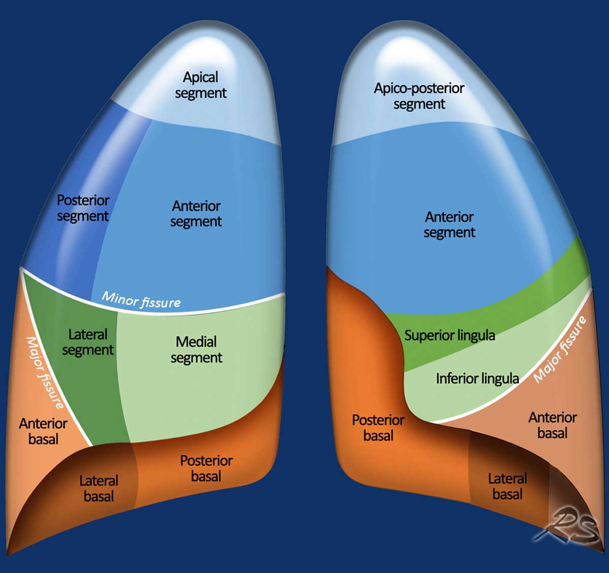
The apical segment of the lung refers to the uppermost functional division of the upper lobe, extending above the clavicle into the root of the neck. Anatomically distinct on the right (segment S1) and left (apicoposterior segment S1+S2), it is prone to specific conditions like apical blebs, Pancoast tumors, and reactivation tuberculosis. Many common myths confuse benign scarring with active disease.
Disclaimer: This article is intended for educational purposes only and should not replace professional medical evaluation, diagnosis, or treatment. Always consult a qualified healthcare provider regarding medical imaging results or persistent chest symptoms.
Understanding the Anatomy of the Apical Lung Segment
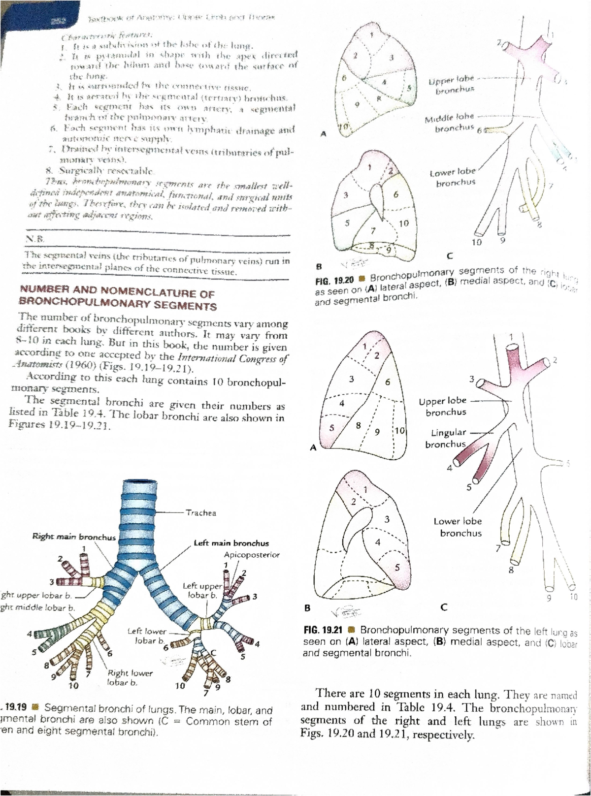
The human lungs are divided into lobes, which are further subdivided into self-contained units known as bronchopulmonary segments. Each bronchopulmonary segment possesses its own segmental bronchus, pulmonary artery branch, and autonomic nerve supply, surrounded by connective tissue septa. Understanding the specific architecture of the apical segment clarifies why certain pulmonary diseases selectively target the top of the lungs.
Structural Anatomy: Right vs. Left Lung
The anatomical configuration of the apex differs between the right and left lungs due to spatial constraints and embryonic development:
- Right Upper Lobe (Apical Segment - S1): The right lung features three distinct lobes. In the right upper lobe, the apical segment (S1) occupies the apex (the highest point). It receives airflow through the apical segmental bronchus ($B^1$), a branch of the right upper lobe main bronchus, and blood flow through the apical branch of the right pulmonary artery.
- Left Upper Lobe (Apicoposterior Segment - S1+S2): The left lung consists of two lobes. Because the cardiac apex occupies space on the left side of the chest cavity, the bronchial tree branches differently. In most individuals, the apical ($B^1$) and posterior ($B^2$) segmental bronchi share a common stem, merging the apical and posterior segments into a single, combined apicoposterior segment (S1+S2).
Positional Anatomy: The Cervical Extension
A common point of confusion in human anatomy involves the physical location of the lung apex relative to the skeletal frame. The apical segment extends roughly 2 to 4 centimeters above the medial third of the clavicle (collarbone) and superior to the first rib.
Because the apical segment projects into the root of the neck (the suprapleural membrane or Sibson's fascia), it sits in close proximity to critical neurovascular structures. These include the subclavian artery and vein, the lower roots of the brachial plexus (C8–T1 nerves), and the cervical sympathetic ganglion trunk. This anatomical relationship explains why disorders originating in the apical segment frequently present with nerve and vascular symptoms rather than typical lower respiratory complaints.
Clinical Significance of the Apical Lung Segment
The physiological environment within the lung apex differs significantly from the base. When an individual stands or sits upright, gravity exerts a major influence on both pulmonary blood flow (perfusion) and ventilation. This unique microenvironment makes the apical segment a target for specific pathological processes.
Top of Lung Apex:
- High Oxygen Tension
- Selective Pathogens (e.g., Reactivation TB)
- Low Blood Perfusion
- Tissue Ischemia & Pleural Thickening
High Oxygen Tension and Reactivation Tuberculosis
In an upright posture, pulmonary perfusion is lowest at the lung apices due to hydrostatic pressure differences across the pulmonary vascular bed. However, ventilation remains relatively high compared to blood flow, resulting in an elevated ventilation-perfusion ratio ($V/Q$).
This high $V/Q$ ratio produces an elevated alveolar partial pressure of oxygen ($PO_2$) in the apical segments. Strictly aerobic pathogens—most notably Mycobacterium tuberculosis—thrive in high-oxygen environments. Consequently, secondary or reactivation tuberculosis disproportionately manifests in the apical and posterior segments of the upper lobes, leading to localized consolidation, tissue necrosis, and cavitary lesions.
Subpleural Blebs, Bullae, and Primary Spontaneous Pneumothorax
The apical segment experiences higher mechanical stretch and mechanical stress than lower parts of the lung due to the effect of gravity pulling the lung downward from the apex. Over time, this chronic mechanical stress can lead to the rupture of elastic fibers in the subpleural tissue, leading to the formation of small air-filled pockets known as blebs (within the visceral pleura) or bullae (within the lung parenchyma).
When an apical bleb ruptures, air leaks directly into the pleural space, collapsing the lung and resulting in a primary spontaneous pneumothorax. This condition predominantly affects young, tall, slender individuals whose thoracic geometry increases the vertical pressure gradient at the lung apex.
Superior Sulcus (Pancoast) Tumors
Neoplasms that develop at the extreme apical boundary of the lung are clinically referred to as superior sulcus tumors, or Pancoast tumors. Most Pancoast tumors are non-small cell lung carcinomas (NSCLCs), predominantly squamous cell carcinomas or adenocarcinomas.
Because of their position at the apical apex, these tumors rarely cause early bronchial obstruction. Instead, they expand locally into the surrounding soft tissues of the thoracic inlet, invading the lower trunks of the brachial plexus, the parietal pleura, upper ribs, and the sympathetic chain.
Debunking 5 Common Myths About the Apical Segment
Misinterpretation of radiological reports and misunderstanding of basic anatomy frequently trigger unnecessary anxiety for patients. Clarifying common misconceptions helps frame medical imaging findings accurately.
| MYTH | REALITY |
|---|---|
| 1. Lungs remain entirely inside the rib cage below the collarbone. | Apex extends 2-4 cm above the clavicle. |
| 2. An "apical shadow" always means active cancer or active tuberculosis. | Most apical caps are benign, age-related. |
| 3. Right and left apical segments are identical. | Left lung combines S1 and S2 into apicoposterior. |
| 4. Apical lung cancer always causes coughing and severe shortness of breath first. | Pancoast tumors present with shoulder/arm pain. |
| 5. Apical blebs only affect chronic smokers. | Tall, thin young adults are at primary risk. |
Myth 1: "The Lungs Stop Below the Collarbone"
The Reality: Many people assume that the collarbone acts as a protective shield covering the absolute top of the lungs. Anatomically, the apex of each lung protrudes above the first rib and medial clavicle into the neck base.
Because the apical segment extends above the clavicle, penetrating trauma to the lower neck or medical procedures such as central venous catheter placement in the subclavian vein carry a risk of accidentally puncturing the apical pleura, potentially causing an iatrogenic pneumothorax.
Myth 2: "An Apical Cap or Shadow Always Indicates Active Malignancy or TB"
The Reality: Seeing terms like "apical capping," "apical pleural thickening," or "apical opacity" on a chest X-ray or CT report often alarms patients. While these findings can represent active disease in appropriate clinical contexts, the vast majority of apical caps are benign.
An apical cap is a localized area of fibrosis and pleural thickening at the lung tip. It affects a large percentage of individuals over age 50 and is typically caused by:
- Chronic low-grade ischemia in the microvascular network of the lung apex.
- Healed, inactive granulomatous infection (such as a resolved prior fungal or mycobacterial exposure).
- Non-specific, age-related scarring from prior local inflammation.
Radiologists distinguish benign apical capping from aggressive lesions by assessing bilateral symmetry, smoothness of borders, absence of underlying bone destruction, and stability across serial imaging studies.
Myth 3: "Right and Left Apical Segments Are Structurally Identical"
The Reality: While the external contour of the lung apices looks symmetrical on chest radiographs, the internal segmental architecture differs. The right lung possesses a standalone apical segment (S1). In contrast, the left upper lobe undergoes a distinct embryonic branching pattern, yielding a combined apicoposterior segment (S1+S2).
This distinction matters during thoracic surgical procedures, such as segmentectomies (removing a single bronchopulmonary segment to preserve healthy tissue). A thoracic surgeon planning a resective surgery must tailor surgical margins and vascular ligations according to these distinct segmental boundaries.
Myth 4: "Apical Lung Cancer Always Causes Classic Respiratory Symptoms First"
The Reality: Primary lung cancers in central airways usually present with persistent cough, hemoptysis (coughing up blood), or wheezing due to airway irritation and blockage. However, tumors in the apical segment (Pancoast tumors) grow in the extreme periphery of the chest wall.
Consequently, patients with apical lung tumors often experience no cough or shortness of breath in the early stages. Instead, they frequently present with neurological and musculoskeletal symptoms caused by local invasion:
- Severe shoulder and arm pain: Caused by tumor invasion into the lower plexus nerves (C8, T1), often radiating down the inner arm to the fourth and fifth fingers.
- Horner’s Syndrome: Caused by disruption of the sympathetic ganglion chain, producing a classic triad of symptoms on the affected side of the face: ptosis (drooping eyelid), miosis (constricted pupil), and anhidrosis (lack of sweating).
- Hand muscle atrophy: Weakness and wasting of the intrinsic muscles of the hand due to nerve entrapment.
Recognizing non-respiratory presentations at the apical segment is critical for avoiding delays in diagnosing superior sulcus neoplasms.
Myth 5: "Apical Blebs Only Affect Older, Heavy Smokers"
The Reality: While chronic obstructive pulmonary disease (COPD) and emphysema produce widespread bullous changes in older smokers, primary spontaneous pneumothorax from apical blebs frequently strikes non-smoking young individuals in their teens, 20s, or 30s.
The predisposing factor for apical blebs in this young demographic is often vertical chest shape. Tall, thin individuals experience a steeper gravitational tension gradient from the base to the top of the lung. This increased tension at the apex leads to micro-tears in the subpleural elastic fibers, forming small isolated blebs that can rupture spontaneously during exercise, air travel, or even while at rest.
Diagnostic Imaging and Evaluation of the Apical Segment
Evaluating the apical segment requires specialized radiological techniques due to overlapping skeletal structures, such as the clavicles, first ribs, and sternoclavicular joints, which can obscure small lesions on standard imaging.
Suspected Apical Pathology:
- Chest X-Ray (Initial screening)
- Apical Lordotic View
- Pleural Thickening
- High-Resolution CT (Detailed characterization)
- Blebs/Bullae Detection
- Pancoast Chest Wall Invasion
- Granuloma vs Active Mass
Chest Radiography (PA and Apical Lordotic Views)
A standard posterior-anterior (PA) chest X-ray is the initial screening tool for evaluating pulmonary symptoms. However, the clavicle and first rib overlap directly on top of the apical segment, creating potential visual blind spots.
To overcome this anatomical overlap, radiologists use an apical lordotic chest X-ray view. In this position, the patient leans backward against the image receptor. This projects the clavicles upward above the lung apices on the film, providing an unobstructed view of the apical segments to clarify questionable shadows seen on standard films.
High-Resolution Computed Tomography (HRCT)
Computed Tomography (CT) is the standard imaging modality for characterizing apical lung lesions. CT scanning provides thin-slice, cross-sectional views that eliminate overlapping bone structures.
- Differentiating Blebs from Parenchyma: HRCT can detect tiny thin-walled subpleural blebs measuring only a few millimeters, which are completely invisible on standard X-rays.
- Characterizing Apical Caps: CT scan imaging clarifies whether an apical cap is a thin, stable fibrotic plaque or a soft-tissue mass requiring biopsy.
- Staging TB and Infections: CT clearly demonstrates apical cavitatory lesions, tree-in-bud nodularity (indicating endobronchial spread), and calcified granulomas.
Magnetic Resonance Imaging (MRI)
While CT is ideal for evaluating lung tissue, Magnetic Resonance Imaging (MRI) is superior for evaluating soft tissue invasion in the apical region. If an apical mass or Pancoast tumor is detected, a thoracic MRI is performed to evaluate whether the tumor has invaded the brachial plexus nerves, subclavian blood vessels, or vertebral bodies.
Comparison Matrix: Apical Segment Pathology vs. Misconceptions
The following matrix contrasts key medical conditions in the apical segment against common clinical misconceptions, detailing the clinical reality and diagnostic evaluation steps.
| Condition / Finding | Common Misconception | Clinical Reality & Pathophysiology | Primary Diagnostic Approach |
|---|---|---|---|
| Benign Apical Cap | Indicates active lung cancer or contagious tuberculosis. | Pleural thickening caused by age-related microvascular ischemia or prior healed inflammation; non-malignant. | Serial Chest CT showing stability; historical comparison. |
| Superior Sulcus (Pancoast) Tumor | Presents primarily with coughing, wheezing, and coughing up blood. | Extrapulmonary compression invading brachial plexus and sympathetic chain; presents with shoulder pain and Horner's syndrome. | Contrast-enhanced CT, Thoracic MRI for plexus involvement, CT-guided biopsy. |
| Apical Subpleural Blebs | Only occurs in elderly individuals with advanced emphysema. | Can form in tall, thin, young non-smokers due to elevated gravitational mechanical strain at the lung apex. | High-Resolution CT (HRCT) of the chest; erect chest radiograph. |
| Reactivation Tuberculosis | Affects all lung segments equally without preference. | Strongly targets apical and posterior segments due to higher regional oxygen tension ($PO_2$) favoring aerobic growth. | Sputum acid-fast bacilli (AFB) smear/culture, chest radiograph, GeneXpert PCR. |
| Anatomical Apex Location | Completely enclosed behind the rib cage below the collarbone. | Projects 2 to 4 cm above the medial clavicle into the root of the neck; accessible above the first rib. | Anatomical examination; ultrasound/CT for procedures near the thoracic inlet. |
When to Seek Medical Evaluation for Apical Segment Symptoms
Incidental imaging findings in the apical segment are common. However, specific progressive or acute symptoms warrant prompt medical evaluation by a healthcare professional.
Red Flag Symptoms
Seek urgent medical evaluation if you experience any of the following symptoms:
- Unexplained, Persistent Shoulder or Upper Back Pain: Severe pain localized to the shoulder girdle, inner scapula, or radiating down the arm (along the C8–T1 nerve distribution), especially when accompanied by weakening hand grip.
- Neurological Changes in the Face (Horner's Syndrome): Unilateral drooping of the eyelid, reduced pupil size in the same eye, or reduced sweating on one side of the face.
- Sudden Sharp Chest Pain and Shortness of Breath: Pleuritic chest pain (worse with deep breathing) localized to one side, which may indicate a spontaneous pneumothorax from a ruptured apical bleb.
- Systemic B-Symptoms: Unexplained fever, night sweats, unintentional weight loss, or persistent cough, which require investigation for pulmonary infections like tuberculosis or fungal granulomatous disease.
Managing Incidental Imaging Reports
If a routine chest X-ray or CT scan reveals an "apical density," "apical pleural cap," or "scarring," maintain a balanced perspective:
- Request Image Comparisons: Ask your physician to compare the new scan with older imaging studies. A finding that has remained unchanged over several years is virtually always benign scarring.
- Avoid Unnecessary Alarm: Recognize that benign pleural capping is a common finding on chest CT scans in adults.
- Follow Recommended Follow-up Intervals: If a nodule or indeterminate opacity is detected, follow the standardized Fleischner Society guidelines for follow-up imaging recommended by your radiologist.
Frequently Asked Questions
What causes an apical cap on a chest X-ray?
An apical cap is usually caused by benign pleural fibrosis resulting from chronic microvascular ischemia at the lung apex, or healed inflammation from a past infection. It is a common finding in older adults and typically does not represent active disease or cancer.
Why does tuberculosis commonly target the apical segment of the lung?
Mycobacterium tuberculosis is an obligate aerobe that thrives in oxygen-rich environments. In an upright individual, the apex of the lung has a high ventilation-perfusion ratio, resulting in higher tissue oxygen tension ($PO_2$) compared to lower lung segments, creating an ideal environment for bacterial growth.
Can an apical lung tumor cause arm pain without a cough?
Yes. Pancoast tumors located at the lung apex grow outward into the surrounding thoracic inlet soft tissues rather than into central airways. They frequently compress the lower roots of the brachial plexus (C8 and T1), causing arm pain, numbness, and hand weakness before any respiratory symptoms occur.
How is a ruptured apical bleb treated?
Treatment depends on the size of the resulting pneumothorax and symptom severity. A small, asymptomatic pneumothorax may be managed conservatively with supplemental oxygen and observation. Larger or symptomatic lung collapses require chest tube insertion to remove pleural air. Recurrent bleb ruptures are often treated with minimally invasive video-assisted thoracoscopic surgery (VATS) to excise blebs (blebectomy) and perform pleurodesis to prevent future recurrences.
How do doctors tell the difference between apical scarring and active cancer?
Radiologists differentiate scarring from malignancy by evaluating imaging characteristics on CT scans. Benign scars typically show smooth, stable borders, bilateral symmetry, calcifications, and no growth over time. Active malignancies often display irregular or spiculated margins, asymmetric growth, invasion into surrounding tissues or bones, and elevated metabolic activity on PET scans.
Practical Recommendations and Next Actions
Understanding the structural and pathological context of the apical segment helps streamline medical decisions and alleviate anxiety over incidental findings. Consider these practical recommendations:
- Review Radiological Findings with Your Physician: If an imaging report notes apical capping or opacities, discuss the findings directly with your doctor. Request a comparison with past imaging to confirm stability.
- Monitor Non-Respiratory Symptoms: Do not ignore localized shoulder, neck, or upper-arm pain that fails to improve with physical therapy, particularly if accompanied by nerve symptoms or drooping eyelids. Prompt evaluation with targeted chest imaging can rule out thoracic outlet or apical lesions.
- Adhere to Smoking Cessation Guidelines: While apical blebs can occur in non-smokers, tobacco and cannabis smoke accelerate tissue degradation, increasing the risk of bullous disease, pneumothorax, and pulmonary neoplasms. Access cessation resources through official health portals like the CDC Smoking and Tobacco Use Resource Center.
- Complete Follow-Up CT Surveillance: If your physician recommends a follow-up chest CT scan in 3, 6, or 12 months for an indeterminate apical nodule, adhere strictly to the schedule to ensure timely evaluation.
Image Gallery
Select an image to open it in a new tab.
Celine Dubois
Contemporary Living and Global Arts Columnist
