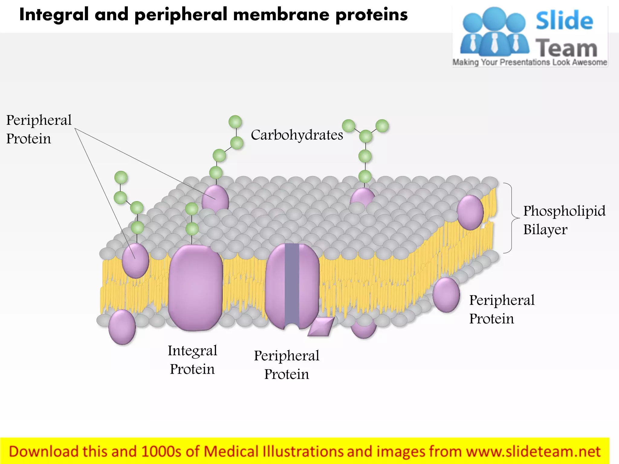
Integral and peripheral proteins are the two primary categories of membrane proteins, classified based on their physical association with the lipid bilayer. Integral proteins span or are deeply embedded within the hydrophobic core of the membrane, whereas peripheral proteins reside on the exterior or interior surfaces, attaching temporarily through weaker molecular interactions.
The Foundation: The Fluid Mosaic Model
To grasp the distinction between these protein types, one must first understand the architecture of the cell membrane. The Fluid Mosaic Model, proposed by S.J. Singer and Garth L. Nicolson in 1972, remains the foundational framework for modern cell biology. The membrane is not a static wall but a dynamic, flexible structure composed of a phospholipid bilayer, cholesterol, carbohydrates, and a complex array of proteins.
The phospholipid bilayer creates a hydrophobic interior—a barrier that prevents water-soluble substances from freely passing through. Proteins within this membrane are essential for transport, cell signaling, enzymatic activity, and structural integrity. Because the environment of the bilayer core is nonpolar, proteins must possess specific structural adaptations to associate with it. These adaptations are the fundamental reason for the divide between integral and peripheral proteins.
Integral Proteins: The Embedded Anchors
Integral proteins, sometimes referred to as intrinsic proteins, are permanently attached to the biological membrane. Their defining characteristic is that they extend into or through the hydrophobic lipid bilayer.
Structural Characteristics
The defining feature of an integral protein is its amphipathic nature. To remain stable within the membrane, the portion of the protein that contacts the fatty acid tails of the phospholipids must be hydrophobic (water-fearing). Conversely, the regions of the protein that protrude into the intracellular cytoplasm or the extracellular matrix are hydrophilic (water-loving).
Many integral proteins are transmembrane proteins, meaning they traverse the entire width of the bilayer. These often feature alpha-helical structures consisting of nonpolar amino acids that anchor them firmly against the lipid tails. Some integral proteins are monotopic, meaning they are associated with only one leaflet of the bilayer but still require the disruption of the membrane to be removed.
Functions of Integral Proteins
Because they are embedded within the membrane, integral proteins are perfectly positioned to mediate interactions between the cell’s internal environment and the outside world.
- Channel and Carrier Proteins: These facilitate the movement of ions, sugars, and amino acids that cannot diffuse through the lipid bilayer.
- Receptor Proteins: These bind to signaling molecules like hormones or neurotransmitters, triggering a cellular response.
- Cell-Cell Adhesion: Integral proteins often link the cytoskeleton of one cell to another or to the extracellular matrix, maintaining tissue cohesion.
Peripheral Proteins: The Transient Surface Associates
Peripheral proteins, or extrinsic proteins, do not penetrate the hydrophobic core of the bilayer. Instead, they are found on the periphery of the membrane, usually attached to the exposed surfaces of integral proteins or the polar head groups of the phospholipids.
Nature of Association
Unlike integral proteins, which require detergents or harsh solvents to be extracted from the membrane, peripheral proteins can typically be removed using milder methods, such as changes in pH or ionic strength. They are held in place by non-covalent interactions, including electrostatic forces and hydrogen bonding.
Because they are not "locked" into the hydrophobic center, peripheral proteins are often more mobile. This allows them to function in temporary capacities, such as signaling cascades that are only activated under specific conditions.
Functions of Peripheral Proteins
Peripheral proteins serve a diverse set of roles that often complement the work of integral proteins:
- Cytoskeletal Anchoring: They often serve as the bridge between the cell membrane and the cytoskeleton, providing shape and structural support.
- Enzymatic Regulation: Many enzymes associated with the membrane are peripheral, allowing them to catalyze reactions near the membrane surface without being permanently tethered to the bilayer structure.
- Signal Transduction: Peripheral proteins often act as secondary messengers in signaling pathways, moving to and from the membrane to propagate signals from receptors to the cell interior.
Comparative Analysis of Membrane Proteins
The differences between these two classes extend beyond their physical location. The following table summarizes the key distinctions to clarify how they function within the cellular architecture.
| Feature | Integral Proteins | Peripheral Proteins |
|---|---|---|
| Membrane Penetration | Extends into or through the bilayer | Sits on the surface only |
| Association Strength | Permanent / Strong | Transient / Weak |
| Extraction Method | Requires detergents to disrupt lipids | Can be removed by pH/salt changes |
| Hydrophobicity | Contains hydrophobic segments | Predominantly hydrophilic |
| Primary Function | Transport, signaling, structure | Signaling, regulation, support |
Methodological Challenges in Research
In 2026, the study of these proteins continues to rely on advanced imaging and biochemical separation techniques. Identifying whether a protein is integral or peripheral is a standard procedure in proteomics.
Researchers typically use "solubilization" experiments. If a sample is treated with a mild salt solution and a protein is released, it is identified as peripheral. If the protein remains attached, researchers introduce detergents—amphipathic molecules that mimic the lipid environment—to dissolve the membrane and isolate the integral protein. Once isolated, researchers use techniques such as Cryo-Electron Microscopy (Cryo-EM) to visualize the protein structure in near-atomic detail, confirming how it integrates with the lipid environment.
Common Misconceptions
One frequent error is the assumption that all membrane proteins are identical in function or that they are all "anchors." In reality, the membrane is a highly specialized environment. Another common mistake is assuming that peripheral proteins are strictly located on the cytoplasmic side of the membrane. In fact, many peripheral proteins are found on the extracellular surface, where they interact with the glycocalyx and play vital roles in immune recognition and cell identification.
Frequently Asked Questions
Can a peripheral protein become an integral protein? No, their classification is based on their amino acid sequence and structural motifs. A protein’s primary structure dictates whether it contains the hydrophobic domains necessary to embed into a lipid bilayer.
Do integral proteins move within the membrane? Yes, according to the Fluid Mosaic Model, both lipids and proteins can move laterally within the plane of the membrane, unless they are anchored to the cytoskeleton or extracellular matrix.
Why is it important to distinguish between these two types of proteins? Understanding this distinction is crucial for pharmacology. Many drugs, such as ion channel blockers, must target specific integral proteins. Knowing if a target is integral or peripheral changes how a drug must be delivered and how it interacts with the cell.
Are peripheral proteins involved in transport? Generally, no. Because they do not span the bilayer, they cannot act as channels or pumps for transporting molecules across the membrane. They are more commonly involved in the regulation of those transport processes.
How does cholesterol affect these proteins? Cholesterol modulates membrane fluidity. By increasing or decreasing the density of the lipid packing, cholesterol can influence how easily integral proteins move or change conformation within the membrane.
Practical Next Steps
If you are analyzing cellular membrane systems, start by verifying the protein's localization using bioinformatics databases such as UniProt. These repositories provide empirical data on whether a protein is classified as a transmembrane (integral) or membrane-associated (peripheral) entity based on its predicted hydropathy plots. By cross-referencing your specific protein of interest with its known domain structure, you can determine the correct experimental approach for isolation and functional study.
Image Gallery
Select an image to open it in a new tab.
Ingrid Bjornson
Global Cyber Policy and Regulation Analyst
