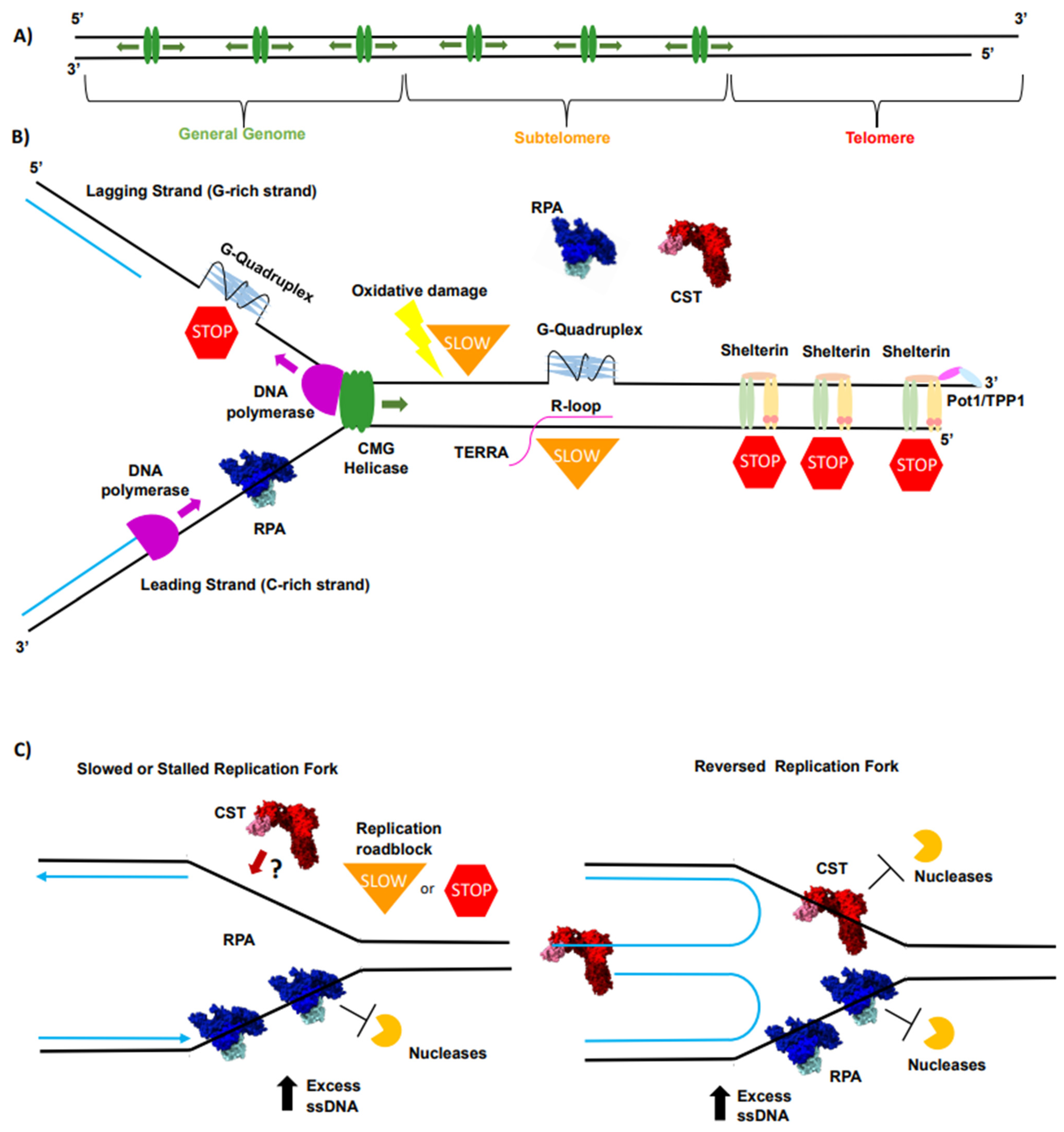
Single-stranded binding proteins (SSBs) protect exposed single-stranded DNA during replication by binding cooperatively to it, preventing secondary structures like hairpins from forming, protecting the strand from nuclease degradation, and recruiting essential replication enzymes to the fork.
Introduction to DNA Replication and the Need for Protection
DNA replication is a fundamental biological process that ensures genetic continuity across cell divisions. In both prokaryotes and eukaryotes, the double helix must be unwound and separated into individual template strands so that replicative DNA polymerases can synthesize complementary new strands. This unwinding is catalyzed by specialized enzymes known as DNA helicases, which disrupt the hydrogen bonds holding the nitrogenous bases together.
However, exposing single-stranded DNA (ssDNA) creates immediate biochemical hazards. Unlike double-stranded DNA (dsDNA), which is structurally stable and protected within its helical conformation, ssDNA is chemically vulnerable, physically flexible, and prone to thermodynamic instability. Without immediate stabilization, exposed ssDNA templates would quickly fold back on themselves, forming complex secondary structures such as hairpins, stem-loops, and cruciforms. These structures present physical roadblocks to the advancing replication machinery, causing polymerases to stall, drop off, or introduce mutations. Furthermore, unprotected ssDNA is an ideal target for cellular nucleases—enzymes designed to degrade stray or damaged nucleic acids.
To solve these challenges, cells employ single-stranded binding proteins. These specialized proteins coat the exposed ssDNA templates immediately behind the advancing helicase, maintaining the strands in an extended, accessible conformation. Understanding how single-stranded binding proteins function requires a detailed examination of their structural biology, cooperative binding mechanisms, and active coordination with the broader replisome.
Structural Characteristics and Biochemical Properties of SSBs
Single-stranded binding proteins are ubiquitous across all domains of life, though their specific structural motifs vary between bacteria, archaea, and eukaryotes. In Escherichia coli, the protein is known simply as SSB and functions as a homotetramer. In eukaryotes, the functional equivalent is Replication Protein A (RPA), a heterotrimer composed of three distinct subunits (RPA70, RPA32, and RPA14). Despite these organizational differences, all single-stranded binding proteins share conserved structural domains optimized for nucleic acid recognition.
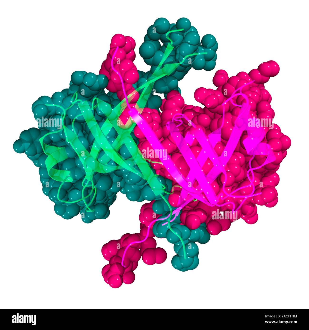
The core functional unit of bacterial SSB is the OB-fold (oligonucleotide/oligosaccharide-binding fold). The OB-fold is a conserved barrel-like structural motif comprising five beta-strands and an alpha-helix. This domain provides a chemical surface rich in aromatic and basic amino acid residues. The aromatic residues—typically tyrosines and phenylalanines—form stacking interactions with the nucleotide bases, while basic residues like lysines and arginines form salt bridges with the negatively charged sugar-phosphate backbone.
| Organism / Complex | Primary Component | Quaternary Structure | Key Nucleic Acid Binding Domain |
|---|---|---|---|
| Escherichia coli | SSB | Homotetramer | OB-fold (Single-stranded DNA-binding domain) |
| Eukaryotes | RPA (Replication Protein A) | Heterotrimer (RPA70, RPA32, RPA14) | Multiple OB-folds across subunits |
| Archaea | RPA-like / SSB | Monomeric or Tetrameric variants | Conserved OB-fold motifs |
The binding mode of bacterial SSB can shift depending on salt concentration and protein-to-DNA ratios, primarily transitioning between a "low-salt" mode (where roughly 65 nucleotides are wrapped around the tetramer) and a "high-salt" mode (where roughly 35 nucleotides are bound). This structural adaptability allows the protein to accommodate varying densities of ssDNA at the replication fork without losing its grip on the template.
Mechanisms of Cooperative Binding and Strand Protection
The primary physical function of single-stranded binding proteins is to coat the unwound template strands uniformly. This is achieved through a phenomenon known as cooperative binding. When a single SSB molecule binds to a stretch of ssDNA, it alters the local conformation and increases the affinity for adjacent SSB molecules to bind immediately next to it. Consequently, SSBs assemble into continuous, tightly packed filaments along the ssDNA template rather than binding randomly or in isolated patches.
This cooperative coating serves several vital protective functions:
- Prevention of Secondary Structures: By neutralizing the base-pairing potential of exposed ssDNA, SSBs prevent intrastrand hydrogen bonding. This keeps the template in an extended, linear state that the DNA polymerase can easily read.
- Defense Against Nucleases: Cellular environments contain numerous single-strand-specific nucleases that can cleave unprotected DNA. The tight protein sheath formed by SSBs physically blocks these enzymes from accessing the phosphodiester backbone.
- Reduction of Thermodynamic Strain: Unwinding dsDNA creates torsional stress. While topoisomerases relieve supercoiling ahead of the fork, SSBs stabilize the localized unwound state, preventing the strands from spontaneously re-annealing behind the helicase.
Importantly, this binding is sequence-independent. SSBs must bind to any sequence of bases without preference, ensuring that the replication machinery does not slow down or detach when encountering adenine-thymine-rich or guanine-cytosine-rich regions. The interaction relies entirely on the universal sugar-phosphate backbone and general stacking with nucleotide bases.
Integration and Coordination Within the Replisome
Single-stranded binding proteins are not passive shields; they are active, dynamic components of the multi-protein replisome. As the replication fork advances, the leading and lagging strands are synthesized simultaneously. The lagging strand, in particular, requires continuous generation of ssDNA loops (Okazaki fragment maturation), creating a high demand for dynamic binding and release of SSBs.
To maintain efficient replication speeds, SSBs interact physically and functionally with various replication enzymes through specific protein-protein interaction domains. These interactions include:
- DNA Helicase: SSBs stimulate the unwinding activity of replicative helicases (such as DnaB in bacteria) by physically interacting with them, ensuring that strand separation and stabilization occur in a coordinated manner.
- DNA Polymerase: SSBs recruit and tether DNA polymerases to the template, increasing the processivity of the enzyme so it can synthesize long stretches of DNA without dissociating.
- Primase and Repair Enzymes: During lagging-strand synthesis, RNA primers must be synthesized repeatedly. SSBs interact with primase (DnaG in bacteria) to regulate primer placement and are subsequently displaced as synthesis proceeds. Furthermore, SSBs recruit various DNA repair and recombination proteins to sites of damaged replication forks.
This network of protein-protein interactions ensures that SSBs are removed efficiently just ahead of the advancing polymerase, preventing steric clashes while maintaining continuous protection of the surrounding ssDNA.
Consequences of SSB Dysfunction
Given the central role of single-stranded binding proteins in maintaining genomic integrity, any defect, mutation, or depletion of these proteins has severe cellular consequences. In experimental models, mutations in the genes encoding SSBs or RPA are frequently lethal or lead to extreme sensitivity to DNA-damaging agents like ultraviolet radiation and chemical mutagens.
When SSBs fail to function correctly, several pathological events occur at the replication fork:
- Fork Stalling and Collapse: Unprotected ssDNA forms aberrant secondary structures, causing DNA polymerases to halt. Prolonged stalling leads to replication fork collapse, double-strand breaks, and subsequent genomic rearrangements.
- Activation of DNA Damage Checkpoints: Unprotected ssDNA is rapidly coated by alternative sensor proteins that trigger DNA damage checkpoints (such as the ATR pathway in eukaryotes), halting the cell cycle to allow for repair.
- Elevated Mutation Rates: Stalled or compromised replication forks often undergo error-prone translesion synthesis or homologous recombination rescue pathways, significantly increasing the frequency of point mutations, deletions, and chromosomal translocations.
Frequently Asked Questions
What is the main difference between bacterial SSB and eukaryotic RPA?
Bacterial SSB is typically a homotetramer composed of four identical subunits containing single OB-folds, whereas eukaryotic Replication Protein A (RPA) is a heterotrimer consisting of three distinct subunits (RPA70, RPA32, and RPA14) that house multiple OB-folds. Despite these architectural differences, both serve the identical core function of protecting and organizing single-stranded DNA during replication.
Why doesn't SSB permanently trap the DNA in a single-stranded state?
SSBs bind DNA through cooperative non-sequence-specific interactions rather than covalent bonds. Their binding is dynamic, and they are actively displaced by DNA polymerases, helicases, and other replisome components as replication proceeds, ensuring the DNA can be successfully converted back into a double helix.
How do single-stranded binding proteins recognize DNA without sequence specificity?
SSBs interact primarily with the sugar-phosphate backbone of the nucleic acid and stack aromatic amino acid residues against the nucleotide bases. Because these structural features are universal to all DNA regardless of sequence, SSBs coat any single-stranded region uniformly.
What happens to SSBs after DNA replication passes through a region?
Once the DNA polymerase synthesizes the complementary strand, the single-stranded template becomes double-stranded once again. The local conformation changes, causing the affinity of the SSB for the nucleic acid to drop, leading to its dissociation and recycling into the nucleoplasm for subsequent binding events.
Conclusion and Further Reading
Single-stranded binding proteins are indispensable guardians of the genome, bridging the gap between physical strand separation and enzymatic synthesis during DNA replication. By preventing secondary structures, shielding vulnerable templates from nucleases, and actively coordinating with the replisome, SSBs ensure the fidelity and speed of genetic duplication. For those seeking a deeper exploration of molecular mechanisms, reviewing structural biology papers on OB-folds and replisome architecture provides valuable insights into cellular preservation of genetic information.
Image Gallery
Select an image to open it in a new tab.

Omar Farooq
EdTech Innovation and Virtual Pedagogy Journalist
