
Deep learning is fundamentally transforming medical diagnostics by enabling automated, high-precision analysis of complex radiological, pathological, and dermatological data. Through multi-layered neural networks, these systems extract subtle pixel-level features imperceptible to the human eye, accelerating disease detection, standardizing interpretation quality, and assisting clinicians in high-stakes medical decision-making.
The Paradigm Shift: From Traditional Imaging to Neural Networks
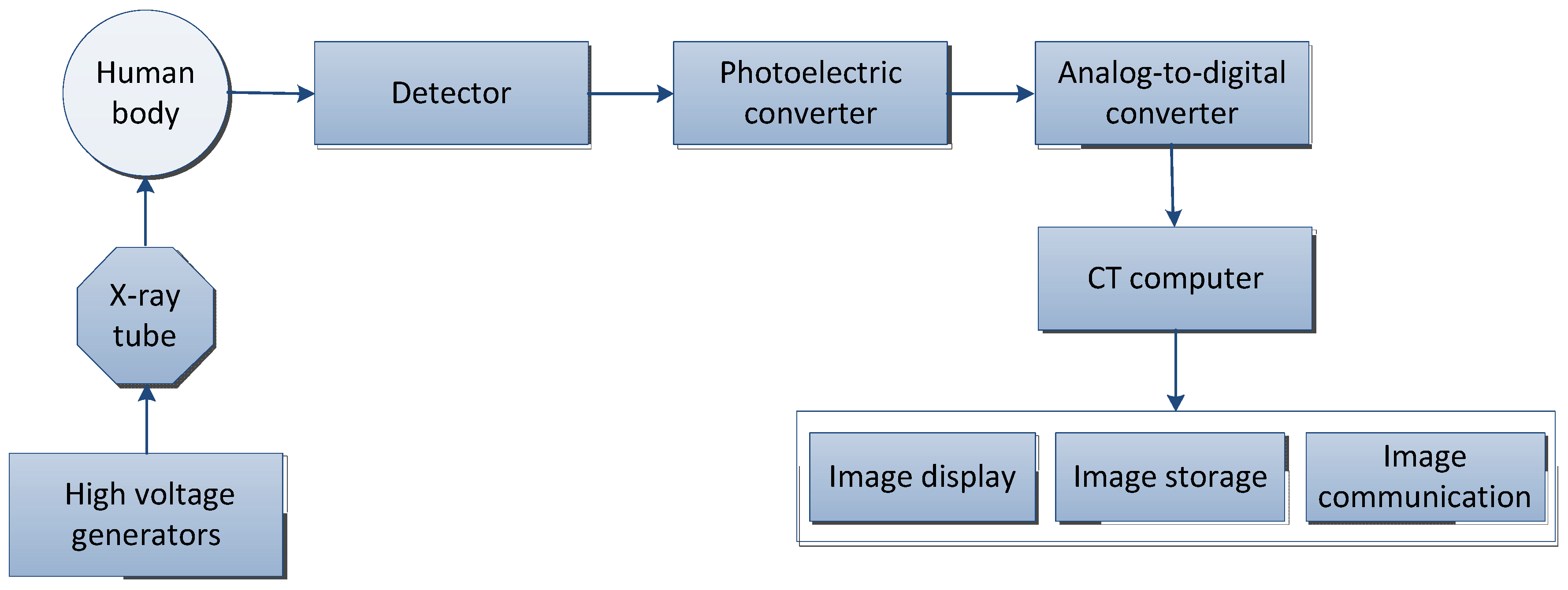
For decades, medical imaging relied exclusively on human visual inspection. Radiologists, pathologists, and cardiologists spent years training to identify structural abnormalities, lesions, and malignancies within computed tomography (CT) scans, magnetic resonance imaging (MRI) outputs, X-rays, and histopathological slides. While this conventional approach remains the gold standard of care, it is intrinsically constrained by human physiological limitations. Fatigue, cognitive overload, inter-observer variability, and sheer volume create bottlenecks in healthcare delivery.
The integration of deep learning—specifically convolutional neural networks (CNNs)—into clinical workflows marks a profound paradigm shift. Instead of relying on hand-crafted rules or static algorithms, deep learning models ingest massive datasets of annotated medical images, autonomously learning hierarchical feature representations. Lower layers of a network might detect basic edges and gradients, while deeper layers synthesize these elements to recognize complex anatomical structures, calcifications, tissue distortions, and early-stage neoplastic growth.
This technological evolution does not seek to replace the clinical workforce; rather, it provides a cognitive force multiplier. By functioning as an automated triage tool, a second reader, or a quantitative measurement engine, deep learning bridges the gap between surging diagnostic volumes and the finite availability of specialized medical professionals.
Core Technical Architectures Driving Diagnostic Innovation
Understanding how deep learning impacts diagnostics requires examining the specific neural network topologies deployed in modern clinical environments. Different medical modalities demand specialized architectural frameworks to process volumetric, spatial, or sequential data effectively.
Convolutional Neural Networks (CNNs)
CNNs serve as the foundational backbone for most medical image analysis tasks. By applying learnable spatial filters across image grids, CNNs preserve local pixel relationships. In chest radiography, models such as ResNet or DenseNet process multi-view scans to flag indicators of pneumonia, pneumothorax, and tuberculosis with sensitivity rates that frequently match or exceed general practitioners.
Vision Transformers (ViTs)
While CNNs excel at capturing local spatial hierarchies, Vision Transformers apply self-attention mechanisms to partition images into smaller patches, evaluating global relationships across the entire scan. In ophthalmology, ViTs are increasingly utilized to analyze retinal fundus photographs, mapping long-range vascular changes associated with diabetic retinopathy and macular degeneration far more effectively than traditional localized filters.
Generative Adversarial Networks (GANs) and Diffusion Models
Generative architectures play a critical role in data augmentation and image reconstruction. Medical datasets are frequently imbalanced, featuring thousands of normal scans but relatively few rare pathologies. GANs synthesize realistic synthetic medical images to train downstream diagnostic classifiers securely without compromising patient privacy. Furthermore, diffusion models accelerate MRI acquisition times by reconstructing high-resolution diagnostic images from sparse, undersampled scan data.

Clinical Applications Across Diagnostic Specialties
The application of deep learning spans virtually every medical specialty that relies on visual data. The following subsections explore how these algorithms revolutionize specific clinical domains.
Radiology and Oncological Screening
In radiology, deep learning excels at early cancer detection. Mammography screening workflows now routinely incorporate deep learning computer-aided detection (CAD) systems. These tools evaluate microcalcifications and architectural distortions in digital breast tomosynthesis, reducing false-positive rates that historically led to unnecessary biopsies and patient anxiety.
Similarly, low-dose CT lung cancer screening programs utilize 3D volumetric CNNs to segment pulmonary nodules. The algorithm measures nodule volume, spiculation, and growth rates across serial scans, providing quantitative risk scores that help thoracic oncologists distinguish benign granulomas from malignant adenocarcinomas.
Pathology and Digital Histopathology
Digital pathology has shifted from glass slides to high-resolution whole-slide imaging (WSI). Because WSIs consist of billions of pixels, manual review is exceptionally time-consuming. Deep learning models segment cellular structures, grade prostate cancer using the Gleason scoring system via Gleason patterns, and quantify mitotic figures in breast cancer biopsies with high reproducibility. This standardization virtually eliminates inter-observer variability between pathologists examining borderline tissue samples.
Cardiology and Echocardiography
Cardiovascular imaging generates complex spatiotemporal data streams. Deep learning algorithms analyze echocardiograms and cardiac MRIs in real-time to automate left ventricular ejection fraction calculation, segment myocardial wall motion abnormalities, and detect early signs of hypertrophic cardiomyopathy. By automating these tedious measurements, technicians can focus more directly on patient acquisition and complex anatomical assessment.
Comparative Analysis of Diagnostic Paradigms
To appreciate the operational impact of integrating deep learning into clinical pipelines, it is helpful to contrast traditional diagnostic workflows with AI-assisted methodologies across critical operational metrics.
| Evaluation Metric | Traditional Diagnostic Workflow | AI-Assisted Deep Learning Workflow |
|---|---|---|
| Primary Bottleneck | Human reading fatigue, high case volumes, and specialist shortages. | Computational infrastructure, algorithm bias, and integration overhead. |
| Processing Speed | Varies by specialist; minutes to hours per complex volumetric scan. | Seconds to minutes for initial triage, segmentation, and scoring. |
| Inter-Observer Variability | Moderate to high, depending on clinician experience and fatigue levels. | Low, ensuring consistent baseline feature extraction across identical inputs. |
| Quantitative Precision | Often relies on manual 2D bounding box measurements and visual estimation. | Automated 3D volumetric segmentation, pixel-level area calculations, and longitudinal tracking. |
| Regulatory Oversight | Standard clinical licensure, institutional peer review, and continuous medical education. | Stringent software-as-a-medical-device (SaMD) clearances (e.g., FDA 510(k), CE mark). |
Implementation Challenges, Regulatory Realities, and Ethical Considerations
Despite profound technical achievements, deploying deep learning models into clinical production environments presents significant structural challenges. Hospitals and healthcare systems must navigate rigorous validation, integration hurdles, and ethical frameworks before clinical deployment.
Dataset Bias and Generalization Failures
A deep learning model is only as unbiased as the data upon which it was trained. If an algorithm is trained primarily on data collected from a single academic medical center utilizing specific imaging hardware, its diagnostic accuracy often plummets when deployed in community hospital settings with different scanner models or diverse patient demographics. Ensuring demographic representation and multi-center external validation is a strict prerequisite for safe clinical translation.
The Black-Box Problem and Explainability
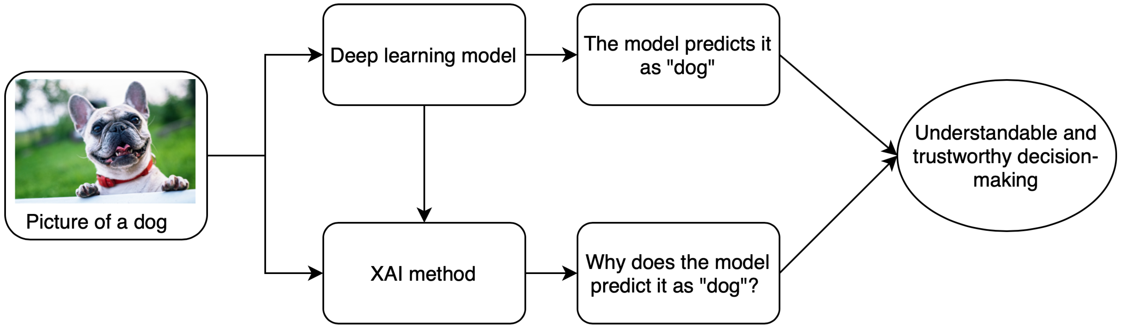
Many complex neural networks operate as "black boxes," producing diagnostic outputs without transparently communicating the physiological rationale behind their predictions. In clinical medicine, accountability is paramount. Clinicians must understand why a model flagged a specific region as malignant before altering a treatment plan. Consequently, the field is rapidly shifting toward Explainable AI (XAI) techniques—such as gradient-weighted class activation mapping (Grad-CAM)—which overlay visual heatmaps onto medical images to highlight the exact pixels that drove the network's decision.
Regulatory Compliance and Software-as-a-Medical-Device (SaMD)
Regulatory bodies, including the United States Food and Drug Administration (FDA) and the European Medicines Agency (EMA), treat clinical deep learning applications as Software-as-a-Medical-Device. Obtaining regulatory clearance requires comprehensive clinical trials demonstrating that the algorithm improves diagnostic accuracy, patient outcomes, or workflow efficiency without introducing unacceptable safety risks. Furthermore, because machine learning models can experience "concept drift" as clinical protocols and imaging hardware evolve, post-market surveillance and continuous performance monitoring are legally mandated.
Best Practices for Clinical Integration and Future Outlook
Successful adoption of deep learning in medical diagnostics requires a structured, multi-disciplinary approach. Healthcare institutions looking to implement these technologies should adhere to established deployment frameworks:
- Conduct Rigorous Local Validation: Never rely solely on vendor-supplied performance metrics. Test models on retrospective local institutional data to measure real-world sensitivity and specificity within your specific patient population.
- Establish Human-in-the-Loop Protocols: Position deep learning tools as decision-support systems rather than autonomous diagnostic agents. Final clinical sign-off and liability must remain with licensed medical professionals.
- Invest in Infrastructure and Interoperability: Ensure that AI diagnostic engines integrate smoothly with existing Picture Archiving and Communication Systems (PACS) and Electronic Health Record (EHR) platforms via standardized protocols like DICOM and HL7/FHIR.
- Prioritize Data Privacy and Security: Implement robust anonymization pipelines, end-to-end encryption, and role-based access controls to protect sensitive patient health information (PHI) during model inference and cloud-based processing.
Looking forward, the convergence of deep learning with multi-omic data—combining medical imaging with genomics, proteomics, and electronic health records—promises to usher in an era of hyper-personalized predictive medicine. By synthesizing macroscopic visual phenotypes with microscopic molecular markers, deep learning will continue to redefine the boundaries of diagnostic precision, ultimately improving patient outcomes on a global scale.
Image Gallery
Select an image to open it in a new tab.



Celine Dubois
Contemporary Living and Global Arts Columnist
