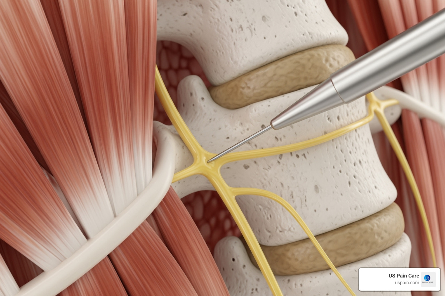
Disc ablation is a minimally invasive medical procedure designed to alleviate chronic lower back pain caused by lumbar discogenic pain, typically by using thermal or radiofrequency energy to disrupt pain-transmitting nerve fibers within or surrounding a damaged spinal disc. This specialized intervention bridges the gap between conservative treatments like physical therapy and invasive open spine surgery, offering targeted relief for carefully selected patients whose discomfort stems from internal disc disruption.
Understanding Discogenic Back Pain and the Role of Ablation
Chronic lower back pain remains one of the leading causes of global disability. A substantial proportion of this pain originates from the intervertebral discs themselves—the shock-absorbing pads situated between the spinal vertebrae. Healthy discs consist of a tough outer ring, the annulus fibrosus, and a gel-like core, the nucleus pulposus. When these structures undergo degenerative changes, annular tears can develop. These fissures allow the inner nuclear material to irritate sensory nerve endings residing within the outer third of the annulus, triggering severe, localized axial back pain that worsens with sitting, bending, or lifting.
Traditional conservative measures—such as non-steroidal anti-inflammatory drugs (NSAIDs), targeted physical therapy, chiropractic care, and epidural steroid injections—frequently fail to resolve discogenic back pain completely. Conversely, major surgical interventions like spinal fusion or total disc replacement carry substantial risks, lengthy recovery periods, and high costs. Disc ablation emerged as an intermediate therapeutic alternative. By selectively targeting and neutralizing the nociceptive (pain-sensing) nerve fibers within the affected disc, ablation aims to reduce or eliminate pain signals transmitted to the brain without compromising the structural integrity or mechanical stability of the spinal motion segment.
Patient Selection and Diagnostic Criteria
Not every patient suffering from lower back pain is a suitable candidate for disc ablation. Strict diagnostic protocols are mandatory to ensure that the pain genuinely originates from the intervertebral disc and not from surrounding structures such as the facet joints, sacroiliac joints, muscular attachments, or neural compression.
Prerequisites for the Procedure
- Chronicity and Failure of Conservative Care: Patients must have experienced chronic lower back pain for a minimum of three to six months, failing to respond adequately to structured non-surgical treatments.
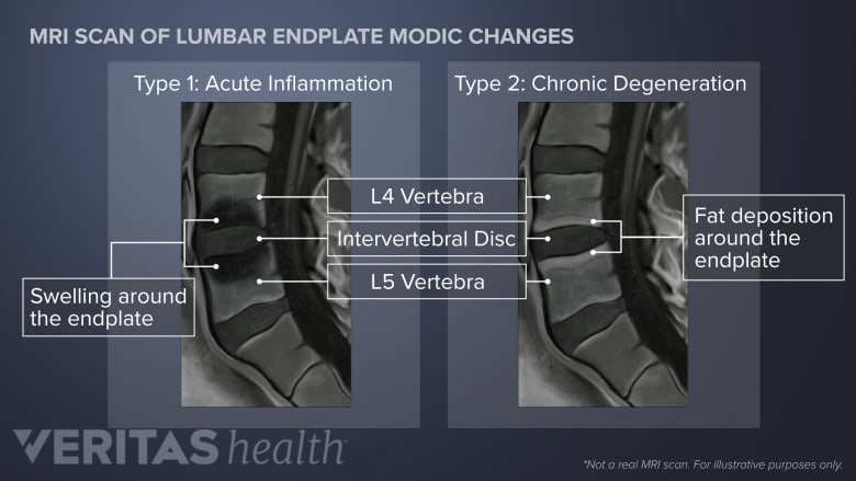
- Imaging Confirmation: Advanced diagnostic imaging, primarily high-resolution Magnetic Resonance Imaging (MRI), must demonstrate signs of degenerative disc disease, such as loss of disc height, hydration loss (dark discs), or annular tears (often visualized as high-intensity zones or HIZs).
- Positive Provocative Discography: Historically, and in many specialized protocols, provocative discography is utilized. During this test, a small amount of contrast dye is injected into the suspected disc under fluoroscopic guidance. If the injection reproduces the patient's exact concordant pain, it strongly implicates that specific disc as the primary pain generator.
- Absence of Severe Pathology: Candidates should not present with severe spinal instability, high-grade spondylolisthesis, significant spinal stenosis, or large herniated discs causing acute motor deficits or cauda equina syndrome, as ablation is ineffective and potentially unsafe for these conditions.
Technical Modalities of Disc Ablation
Several medical device technologies and techniques exist to perform disc ablation. While they share the common goal of disrupting nerve transmission within or adjacent to the disc, the delivery mechanisms and energy sources differ significantly.
Intradiscal Electrothermal Therapy (IDET)
IDET was one of the earliest and most studied thermal annular procedures. During IDET, a flexible catheter with a heating element is guided via a needle into the posterior aspect of the annulus fibrosus under real-time fluoroscopic visualization. The catheter is heated to a controlled high temperature for a specified duration. This thermal energy aims to denature the nociceptive nerve fibers embedded in the annular fissures and cause collagen remodeling to seal small tears in the disc wall.
Radiofrequency Ablation (RFA) and Bicongular / Cooled RFA
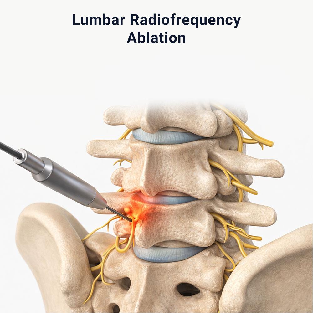
Radiofrequency ablation utilizes alternating current at high frequencies to generate heat, which is then applied to target nerves. In the context of disc-related pain, RFA may be directed at the basivertebral nerve (the nerve supplying the vertebral body endplates, targeted via procedures such as Intracept) or applied directly to the annular branches. Cooled radiofrequency ablation allows for a larger spherical lesion zone, increasing the probability of successfully interrupting pain signals from complex neural networks surrounding degenerated discs.
Laser Disc Decompression and Ablation
Laser-assisted procedures utilize laser energy delivered through a microscopic fiber-optic cable inserted into the disc. This energy can simultaneously vaporize a small portion of the nucleus pulposus to reduce intradiscal pressure (decompression) and coagulate localized pain receptors in the surrounding tissue.
Step-by-Step Overview of the Procedure
Understanding the clinical workflow helps demystify the intervention and prepares patients for what to expect before, during, and immediately after the procedure.
Patient Evaluation & MRI/Discography
Pre-Procedure Preparation & Local Anesthesia
Fluoroscopic Needle Placement into Target Disc
Delivery of Thermal/RF Energy to Target Nerves
Post-Procedure Monitoring & Discharge Home
Pre-Procedural Preparation: Patients are instructed to fast for a brief period before the procedure. Anticoagulant medications (blood thinners) must be temporarily discontinued under the supervision of the prescribing physician to minimize bleeding risks.
Anesthesia and Positioning: The procedure is typically performed in an outpatient surgical suite or hospital radiology department. The patient lies prone (face down) on a specialized fluoroscopy table. Local anesthesia is administered to numb the skin and subcutaneous tissue along the needle insertion pathway. Mild intravenous sedation is often provided to ensure patient comfort, though general anesthesia is rarely required.
** Needle Insertion and Guidance:** Utilizing bi-planar fluoroscopy (X-ray guidance), the physician carefully advances a specialized introducer needle through the skin and soft tissues, past the nerve roots, and directly into the target intervertebral disc or annular fissure.
Energy Delivery: Once the precise positioning is verified via contrast medium and imaging, the ablation catheter, radiofrequency probe, or laser fiber is deployed. Thermal or radiofrequency energy is applied according to a calibrated protocol (often lasting between 10 to 20 minutes depending on the specific technology used).
Completion and Recovery: The device and needle are removed, and a sterile bandage is applied. The patient is transferred to a recovery area for monitoring over one to two hours before being discharged into the care of a designated driver.
Comparative Overview of Interventional Back Pain Treatments
To contextualize disc ablation, it helps to examine how it compares with alternative interventional modalities for chronic lower back pain.
| Treatment Modality | Primary Mechanism | Invasiveness | Typical Recovery Time | Target Pathology |
|---|---|---|---|---|
| Epidural Steroid Injection | Anti-inflammatory medication delivered to the epidural space | Minimally Invasive (Injections) | 1 to 3 days | Radicular pain, nerve root impingement, herniated discs |
| Disc Ablation (Thermal/RF) | Thermal destruction of nociceptive nerve fibers in/around the disc | Minimally Invasive (Percutaneous) | Few days to 2 weeks | Discogenic back pain, annular tears, internal disc disruption |
| Microdiscectomy | Surgical excision of herniated disc material pressing on nerves | Moderately Invasive (Open/Tubular) | 4 to 6 weeks | Symptomatic disc herniation with radiculopathy |
| Lumbar Spinal Fusion | Permanent stabilization and bony fusion of adjacent vertebrae | Highly Invasive (Open Surgery) | 3 to 6 months | Spinal instability, severe spondylolisthesis, advanced degeneration |
Post-Procedural Recovery and Rehabilitation
Recovery following disc ablation is generally rapid compared to open spinal surgery, but it requires adherence to specific physical restrictions to allow the treated tissues to heal.
- Immediate Post-Op Days: Mild to moderate soreness at the needle insertion site and a temporary flare-up of back spasms are common during the first week. This is managed with prescribed analgesics, muscle relaxants, and cold therapy.
- Activity Modifications: For the first two to four weeks, patients are advised to avoid heavy lifting (typically nothing over 10–15 pounds), excessive spinal flexion (bending forward), twisting motions, and prolonged sitting.
- Physical Therapy Integration: As acute post-procedural soreness subsides, structured physical therapy is introduced. Core stabilization, lumbar flexibility exercises, and postural re-education are essential to restore muscular support to the spine and prevent recurrence of pain.
- Timeline for Pain Relief: Unlike surgical interventions that provide immediate mechanical relief, nerve ablation relies on physiological healing and gradual neural desensitization. Significant pain reduction often manifests progressively over 6 to 12 weeks following the procedure.
Risks, Complications, and Limitations
While disc ablation is considered safe when performed by experienced interventional spine specialists or neurosurgeons, it carries inherent medical risks. Patients must weigh these potential complications against the expected benefits.
- Infection: As with any percutaneous procedure, there is a minor risk of intradiscal infection (discitis), which can cause severe pain and requires antibiotic therapy or surgical washout.
- Nerve Injury: Misplacement of the needle or thermal spread can theoretically cause temporary or permanent injury to adjacent spinal nerves or the spinal cord, resulting in numbness, weakness, or neuropathic pain.
- Increased Pain or Lack of Efficacy: In some cases, the procedure fails to provide meaningful relief, and a small percentage of patients experience transient or prolonged worsening of back pain due to thermal irritation of surrounding tissues.
- Structural Weakness: Excessive thermal energy applied to the disc can theoretically compromise the structural integrity of the annulus fibrosus, though modern procedural guidelines are carefully calibrated to avoid weakening the disc wall.
Frequently Asked Questions
How long does pain relief from disc ablation last?
The duration of pain relief varies considerably among individuals. Successful procedures can provide pain reduction lasting from several months to multiple years. Because intervertebral discs continue to age and degenerate over time, ablation does not halt the underlying degenerative process, meaning pain may eventually return if structural stresses persist.
Is disc ablation covered by health insurance?
Insurance coverage for disc ablation varies widely depending on the specific technology used, the policy provider, and strict adherence to pre-authorization criteria. Many insurers classify certain intradiscal therapies as investigational or require extensive documentation proving that conservative measures and diagnostic discography have failed. Patients must verify coverage details with their insurance provider prior to scheduling the procedure.
Can disc ablation be repeated if the pain returns?
In select cases where the procedure initially provided substantial relief that subsequently waned, a repeat ablation may be considered. However, this decision depends heavily on repeat imaging, the condition of the remaining disc structure, and the safety of re-accessing the previously treated spinal level.
Will disc ablation completely cure my degenerative disc disease?
No. Disc ablation is purely a palliative and pain-mitigating intervention, not a cure for degenerative disc disease. It is designed to interrupt pain signals originating from a specific damaged disc, but it does not regenerate healthy disc tissue or reverse structural degeneration.
What is the difference between medial branch block and disc ablation?
A medial branch block (and subsequent facet radiofrequency ablation) targets the facet joints located at the back of the spine, which cause arthritic back pain. Disc ablation targets the interior or outer wall of the intervertebral disc itself, which generates axial pain due to internal disruption and annular tears. They treat entirely different anatomical sources of back pain.
Practical Next Steps for Patients
Patients experiencing persistent lower back pain who suspect a discogenic origin should initiate a comprehensive consultation with a board-certified spine specialist, physiatrist, or interventional pain physician. Bringing a complete history of previous diagnostic imaging (such as MRI reports), physical therapy notes, and medication trials will streamline the evaluation process. Ensuring that all non-invasive options have been thoroughly explored is the most critical prerequisite before pursuing advanced percutaneous interventions like disc ablation.
Image Gallery
Select an image to open it in a new tab.



Lars Thomsen
Global Labor Markets and Economic Strategy Editor
