Understanding Gram-Positive Bacteria in Pairs: Clinical Significance and Identifying Pathogens

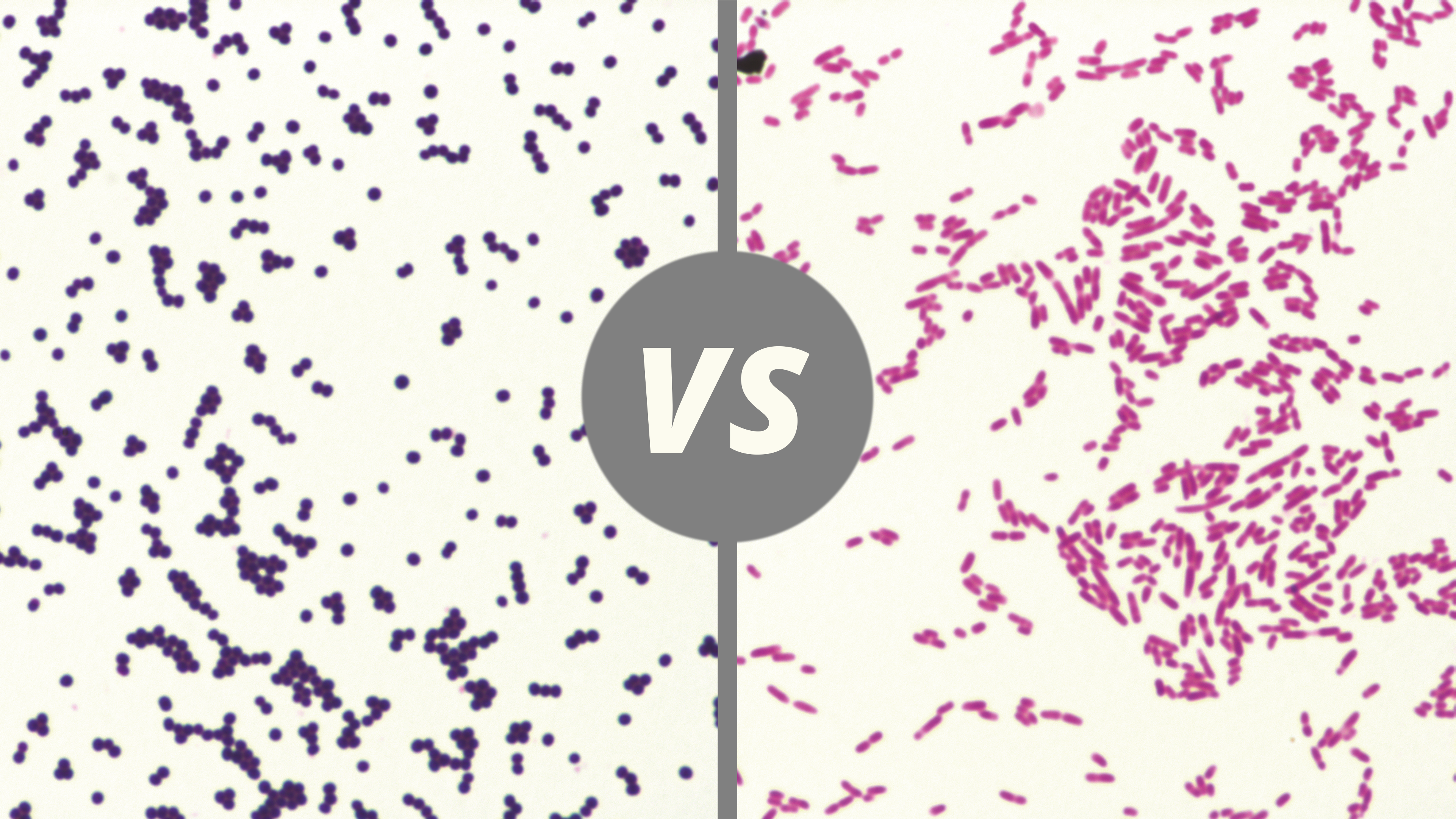
A finding of "Gram-positive bacteria in pairs" on a laboratory report describes spherical bacteria (cocci) that retain purple dye during staining and appear coupled together. Most commonly, this morphology indicates Streptococcus pneumoniae, a major cause of pneumonia and meningitis, though it can also represent Enterococci or other species requiring clinical correlation.
The Gram stain remains one of the most critical preliminary tests in microbiology. When a clinician receives a preliminary report stating "Gram-positive cocci in pairs" (often abbreviated as GPC in pairs or diplococci), it provides an immediate clue toward the identity of the invading pathogen. In 2026, while rapid molecular diagnostics have become more prevalent, the visual confirmation provided by a Gram stain remains a gold standard for initial bedside decision-making and empiric antibiotic selection.
The Science of the Gram Stain and Bacterial Morphology
To understand what "Gram-positive in pairs" means, one must first understand the laboratory process that produces this description. Developed in 1884 by Hans Christian Gram, the Gram stain categorizes bacteria based on the physical and chemical properties of their cell walls.
The Gram-Positive Cell Wall
Gram-positive bacteria possess a thick layer of peptidoglycan, a polymer consisting of sugars and amino acids that forms a mesh-like layer outside the plasma membrane. This thick layer (ranging from 20 to 80 nanometers) acts as a trap for the primary stain used in the process, crystal violet.
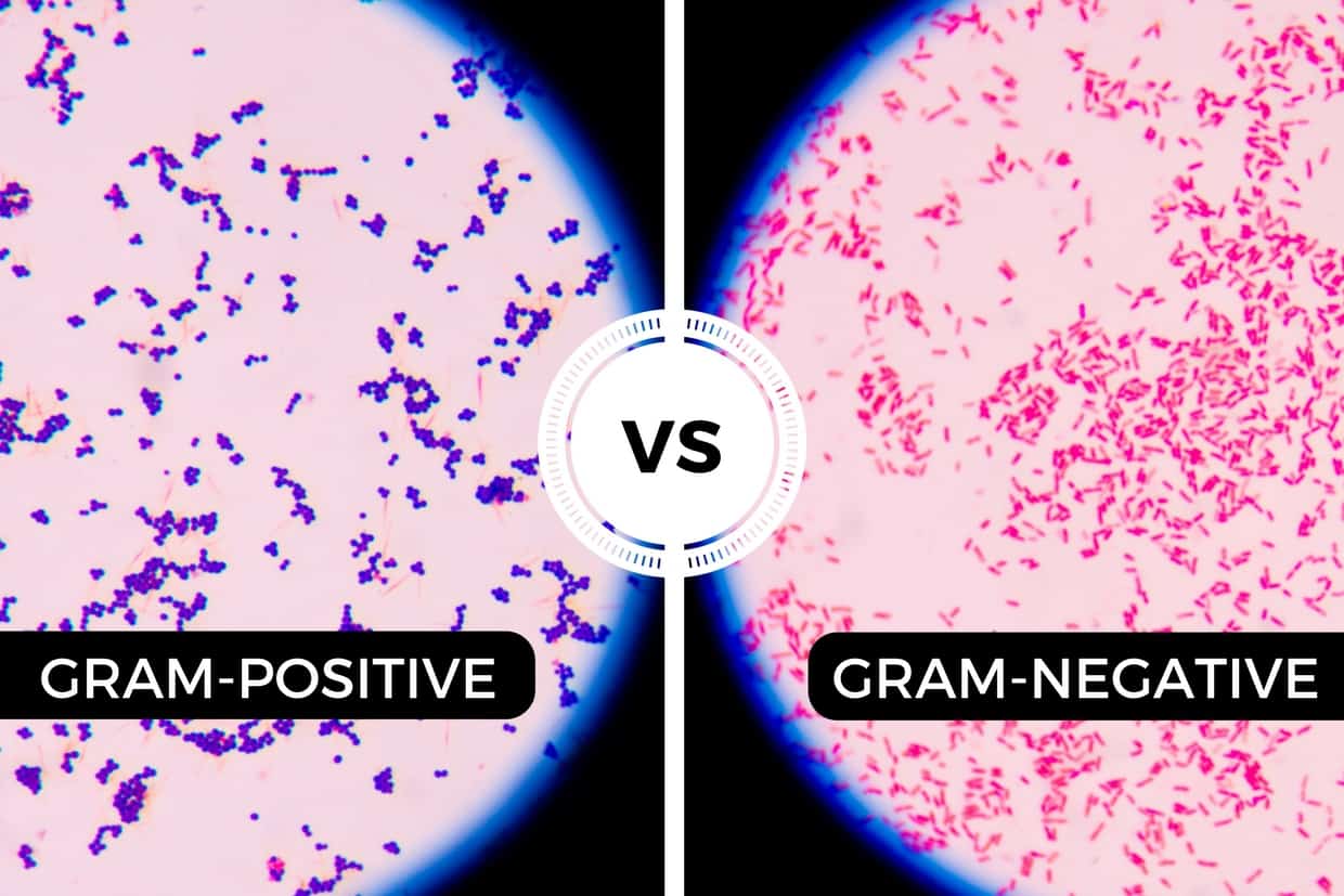
When iodine is added as a mordant, it forms a large crystal violet-iodine complex within the peptidoglycan. Even when a decolorizer (alcohol or acetone) is applied, the thick cell wall of a Gram-positive organism does not shed the purple dye. Consequently, these bacteria appear deep blue or purple under a light microscope.
Defining "In Pairs" (Diplococci)
The term "in pairs" refers to the cellular arrangement. Bacteria divide through binary fission. In some species, the daughter cells remain attached to one another after division rather than fully separating or forming long chains or clusters.
When cocci (spherical bacteria) remain in pairs, they are technically termed diplococci. This specific arrangement is a vital diagnostic marker. While many bacteria can appear as individual spheres, only a select few pathogenic groups consistently present in this "paired" configuration.
Primary Pathogens: What "In Pairs" Usually Means
When a laboratory technician identifies Gram-positive cocci in pairs, they are typically looking at one of several clinically significant organisms. The exact identity often depends on the source of the sample (e.g., blood, sputum, or cerebrospinal fluid).
Streptococcus pneumoniae (Pneumococcus)
This is the most common and significant organism associated with the description "Gram-positive diplococci." Under the microscope, S. pneumoniae often exhibits a very specific "lancet-shaped" appearance—meaning the pairs are slightly pointed at the ends rather than perfectly round.
- Clinical Relevance: It is a leading cause of community-acquired pneumonia, bacterial meningitis, otitis media (ear infections), and sinusitis.
- Identification Clues: In 2026, clinicians look for the presence of a capsule around the pairs, which may appear as a "halo" or clear zone around the purple cells in certain staining conditions.
Enterococcus Species
Enterococcus faecalis and Enterococcus faecium are Gram-positive cocci that frequently appear in pairs and short chains. Unlike the lancet-shaped pneumococcus, enterococci tend to be more oval or egg-shaped.
- Clinical Relevance: These are common causes of urinary tract infections (UTIs), endocarditis, and intra-abdominal infections. They are particularly notorious in hospital settings due to their intrinsic resistance to many antibiotics and the rise of Vancomycin-Resistant Enterococcus (VRE).
- Identification Clues: They are often found in samples derived from the GI tract or catheterized urine.
Staphylococcus aureus (Early Division)
While Staphylococcus species are classically known for appearing in "grape-like clusters," they can occasionally appear in pairs during the early stages of rapid multiplication. However, a seasoned microbiologist can usually distinguish these from true diplococci by observing the surrounding field for the characteristic cluster formations.
Clinical Scenarios and Diagnostic Importance
The presence of Gram-positive pairs is not just a biological curiosity; it dictates the immediate course of medical treatment. Because cultures can take 24 to 48 hours to grow, the Gram stain result serves as the "early warning system."
Sputum Samples and Pneumonia
In a patient with a cough, fever, and chest X-ray showing an infiltrate, a sputum Gram stain showing "many Gram-positive cocci in pairs" suggests a diagnosis of pneumococcal pneumonia. If the sample also shows many neutrophils (white blood cells), the likelihood of an active infection is very high.
Cerebrospinal Fluid (CSF) and Meningitis
This is perhaps the most critical scenario. If Gram-positive diplococci are seen in the CSF of a patient with a stiff neck and altered mental status, it is a medical emergency. Streptococcus pneumoniae is a common cause of adult bacterial meningitis, and identifying it in pairs allows doctors to immediately start high-dose targeted antibiotics like ceftriaxone and vancomycin.
Blood Cultures and Sepsis
When a blood culture bottle "flags" as positive, the first thing a lab does is a Gram stain. Seeing pairs helps narrow down the source of the sepsis. For instance, pairs in the blood might suggest a primary lung infection that has spread to the bloodstream.
Comparative Morphology of Gram-Positive Cocci
To better understand how "pairs" differ from other arrangements, the following table compares common Gram-positive cocci.
| Arrangement | Description | Common Examples | Typical Infections |
|---|---|---|---|
| Pairs (Diplococci) | Two spherical cells joined together; often lancet-shaped. | Streptococcus pneumoniae | Pneumonia, Meningitis, Sinusitis |
| Chains | Spherical cells linked in a linear fashion like a string of beads. | Streptococcus pyogenes (Group A) | Strep throat, Scarlet fever, Cellulitis |
| Clusters | Spheres arranged in irregular, grape-like bunches. | Staphylococcus aureus | Skin abscesses, MRSA, Bone infections |
| Tetrads | Groups of four cocci arranged in a square. | Micrococcus luteus | Usually skin flora (rarely pathogenic) |
The Role of Modern Technology in 2026
While the visual identification of Gram-positive pairs remains foundational, 2026 healthcare utilizes integrated diagnostic pathways. Once pairs are identified, laboratories often reflexively perform rapid tests to confirm the species and detect resistance markers.
- MALDI-TOF MS: Matrix-Assisted Laser Desorption/Ionization-Time of Flight Mass Spectrometry. This technology identifies the protein "fingerprint" of the bacteria. If a lab sees pairs on a slide, they can often get a definitive species identification within minutes of a positive culture using this method.
- Rapid PCR Panels: These molecular tests look for the DNA of S. pneumoniae or Enterococcus directly from the sample. Even if the bacteria are dead (perhaps because the patient already started antibiotics), the DNA can still be detected.
- Automated Sensitivity Testing: Once the organism in pairs is grown, automated systems determine which antibiotics will be most effective, a process that has become significantly faster and more precise by 2026.
Limitations and Potential Misinterpretations
It is important to note that a Gram stain is an interpretation by a human or an AI-assisted imaging system. Several factors can lead to confusion regarding "Gram-positive in pairs."
- Decolorization Errors: If the decolorizer is left on too long, Gram-positive bacteria can lose their purple stain and appear pink (Gram-negative). This might lead a clinician to mistakenly look for Neisseria (Gram-negative diplococci) instead of S. pneumoniae.
- Sample Contamination: Many "Gram-positive cocci in pairs" are normal flora. For example, Viridans group streptococci are normal inhabitants of the mouth and can appear in pairs in a sputum sample that was not collected properly.
- Antibiotic Pre-treatment: If a patient has already taken antibiotics, the bacteria may become "stressed." This can cause them to lose their typical shape or staining characteristics, making pairs harder to identify.
Medical Disclaimer
The information provided in this article is for educational and informational purposes only and is not intended as medical advice. Laboratory results should always be interpreted by a qualified healthcare professional in the context of a full clinical evaluation. If you or someone you know is experiencing symptoms of an infection, seek medical attention immediately.
Frequently Asked Questions
Is "Gram-positive in pairs" always a sign of infection?
Not necessarily. The significance of the finding depends entirely on where the sample was taken. If found in a sterile site like the blood or spinal fluid, it almost always indicates a serious infection. However, if found in a throat swab or a non-sterile skin sample, it may simply represent "colonization" or normal bacteria that live on the body without causing harm.
How is an infection caused by Gram-positive pairs treated?
Treatment depends on the specific species identified and its antibiotic sensitivity. Streptococcus pneumoniae is often treated with penicillins, cephalosporins (like ceftriaxone), or macrolides, though resistance is a growing concern. Enterococci are more difficult to treat and may require specialized antibiotics like vancomycin, linezolid, or daptomycin.
What is the difference between "pairs" and "chains"?
"Pairs" refers specifically to two cells joined together. "Chains" refers to longer strings of cells. While both are characteristic of different types of Streptococcus, S. pneumoniae is the classic "pair" former, whereas S. pyogenes (the cause of Strep throat) is the classic "chain" former. This distinction helps the lab narrow down the species before the final culture is ready.
Can Gram-positive pairs be seen without a microscope?
No. Individual bacteria are far too small to be seen with the naked eye. Even the "pairs" arrangement requires a light microscope with high magnification (usually 1000x with oil immersion) to be visualized by a technician or pathologist.
Why do some reports say "lancet-shaped diplococci"?
"Lancet-shaped" is a descriptive term used specifically for Streptococcus pneumoniae. It means the cocci are not perfectly round but are slightly elongated with a pointed end, resembling the blade of a lancet. This specific morphology is highly suggestive of this particular pathogen.
Recommendations for Patients and Caregivers
If you are reviewing a lab report that mentions "Gram-positive cocci in pairs," the most important next step is to consult with the ordering physician. This finding is a preliminary result, often called a "preliminary microbiology report."
- Ask for the Source: Clarify if the bacteria were found in a sterile site (like blood) or a site with normal bacteria (like the mouth).
- Inquire About Targeted Therapy: Ask if the current antibiotic regimen covers Streptococcus pneumoniae or Enterococcus, as these are the most likely suspects.
- Monitor Symptoms: If this finding was from a sputum sample, watch for worsening shortness of breath or high fevers. If it was from a CSF sample, monitor for changes in mental clarity or severe headache.
- Wait for the Final Culture: Remember that the Gram stain is just the first step. The "Final Culture and Sensitivity" report, usually available within 48-72 hours, will provide the definitive name of the bacteria and exactly which medicines will kill it.
Image Gallery
Select an image to open it in a new tab.


Celine Dubois
Contemporary Living and Global Arts Columnist
