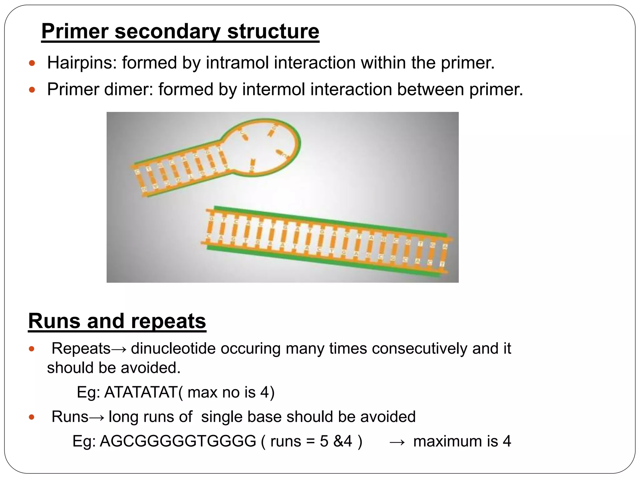
Primer dimers are non-specific PCR artifacts formed when primer molecules hybridize to each other rather than the target DNA, subsequently becoming extended by DNA polymerase. These small, double-stranded products compete for reaction components, significantly reducing amplification efficiency and complicating quantification in downstream applications like quantitative PCR (qPCR) or next-generation sequencing (NGS).
The Nature of Primer Dimers
In the field of molecular biology, the Polymerase Chain Reaction (PCR) remains the gold standard for DNA amplification. However, even in 2026, the formation of primer dimers (PDs) continues to be one of the most persistent challenges for researchers. A primer dimer is essentially a byproduct of the PCR process where two primer molecules—either the same species (self-dimers) or different species (cross-dimers)—anneal to one another via short regions of complementarity.
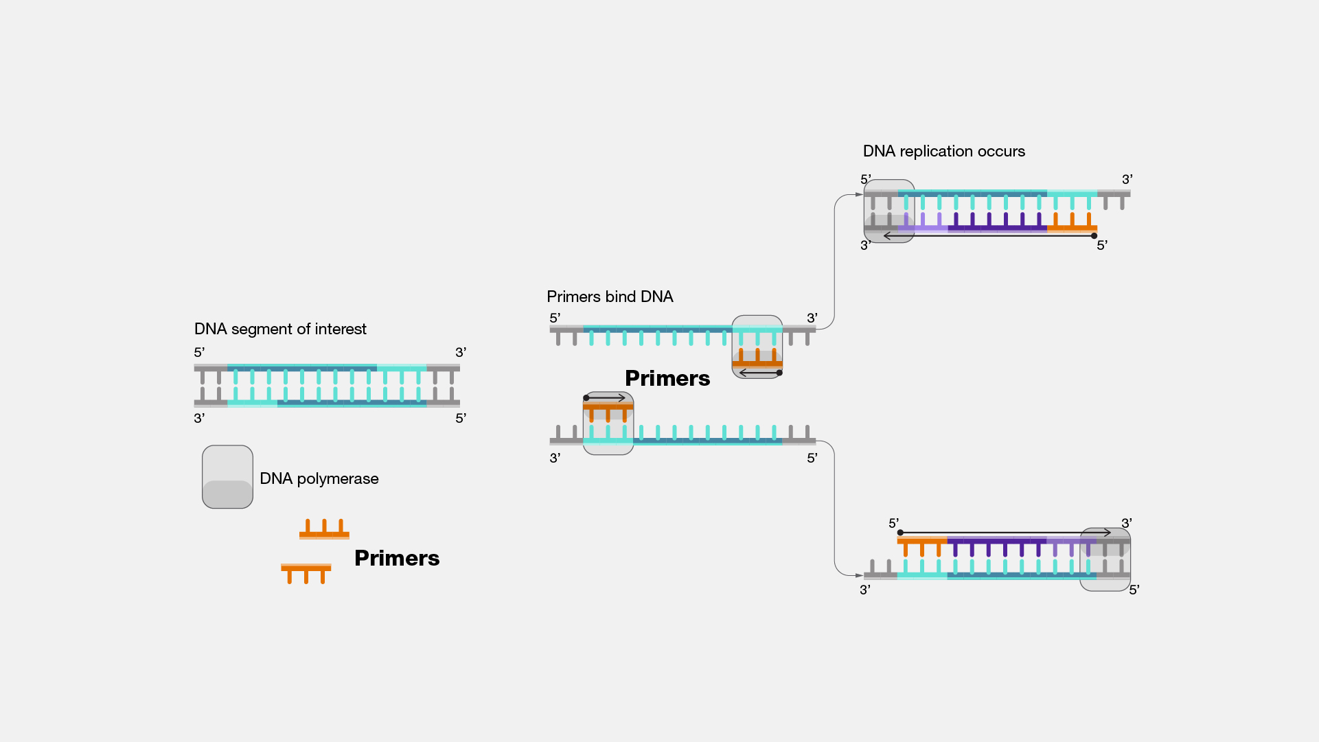
The problem begins during the initial stages of the reaction, often while the master mix is being prepared at room temperature. If the 3' ends of two primers share even a few complementary bases, DNA polymerase can recognize this as a template-primer complex. Once the polymerase extends these primers, the resulting product is a short, double-stranded DNA fragment that contains the sequences of both primers. Because this "dimer" now possesses perfect complementarity to the primers used in the reaction, it is amplified with extremely high efficiency in every subsequent cycle, often outcompeting the intended target sequence.
The consequences of primer dimer formation are twofold. First, they consume critical reagents, including deoxynucleotide triphosphates (dNTPs), primers, and the DNA polymerase enzyme itself. Second, in fluorescence-based detection methods like SYBR Green qPCR, primer dimers produce a signal that is indistinguishable from the target signal, leading to overestimation of DNA concentration or false-positive results in diagnostic assays.
Mechanisms of Formation: Why Primers Bind Together
The formation of primer dimers is governed by the laws of thermodynamics and the kinetic properties of DNA polymerase. Understanding these mechanisms is the first step toward preventing them.
Sequence Complementarity and the 3' End
The most critical factor in PD formation is the sequence at the 3' terminus of the primers. DNA polymerase adds nucleotides only to the 3' hydroxyl group. Therefore, if two primers hybridize in a way that their 3' ends are "pointing" toward each other with even two or three matching base pairs, the polymerase will lock onto this structure and begin extension. This is often referred to as "3' end stability." While internal complementarity can cause secondary structures like hairpins, it is the 3' complementarity that directly facilitates the creation of the extendable dimer.
Thermodynamics and Gibbs Free Energy ($\Delta G$)
In 2026, sophisticated primer design software focuses heavily on the Gibbs Free Energy ($\Delta G$) of potential dimer structures. $\Delta G$ represents the amount of energy released or consumed during the hybridization process. A negative $\Delta G$ indicates a spontaneous reaction. For primer dimers, a $\Delta G$ value more negative than -9 kcal/mol is generally considered a high risk for significant artifact formation. Modern laboratory protocols prioritize primers with the highest possible (least negative) $\Delta G$ for self- and cross-dimerization to ensure reaction cleanliness.
The Competition Effect
A fundamental principle of PCR kinetics is that shorter sequences are amplified more efficiently than longer ones. Because primer dimers are typically only 30 to 50 base pairs long, they denature quickly and anneal almost instantaneously. In a reaction where the target sequence is several hundred base pairs long, the primer dimers can go through many more "effective" cycles of replication in the same timeframe, eventually exhausting the reaction's resources before the target has reached detectable levels.
How to Detect Primer Dimers in the Laboratory
Detection is straightforward but requires careful analysis of the data produced during and after the PCR run. Relying solely on the presence of a "band" or a "signal" is insufficient for professional-grade molecular diagnostics.
Gel Electrophoresis
The most traditional method for detecting primer dimers is agarose gel electrophoresis. When a PCR product is run on a gel (typically 2% to 3% agarose for better resolution of small fragments), primer dimers appear as a "fuzzy" or "smeary" band at the very bottom of the lane, usually below the 50 bp marker. If the target band is faint or absent while the dimer band is intense, it indicates that the dimers have successfully outcompeted the target.
Melting Curve Analysis in qPCR
In quantitative PCR using intercalating dyes like SYBR Green, melting curve analysis is the primary tool for distinguishing between the target and artifacts. After the final amplification cycle, the thermal cycler gradually increases the temperature while monitoring fluorescence. Because primer dimers are short, they have a lower melting temperature ($T_m$) than the longer target product.
A typical melting curve with primer dimers will show two distinct peaks:
- A primary peak at a higher temperature representing the target DNA.
- A secondary, smaller peak at a lower temperature (often between 65°C and 75°C) representing the primer dimers.
If the "No Template Control" (NTC) shows a peak in this lower temperature range, it is a definitive sign of primer dimer formation, as there is no template DNA present to serve as a substrate.
Cq Value Shifts
In qPCR, the quantification cycle ($C_q$) is the point at which the fluorescence signal crosses the threshold. Significant primer dimer formation can cause a "leftward shift" in the $C_q$ (making it appear as if there is more template than there actually is) or, conversely, a "rightward shift" if the dimers are so prevalent that they inhibit the early-stage amplification of the target.
Strategic Mitigation: Reducing and Eliminating Dimers
Eliminating primer dimers requires a multi-faceted approach involving software-driven design, chemical additives, and thermal cycling adjustments.
Advanced Primer Design
The first line of defense is the use of robust design tools such as NCBI Primer-BLAST or specialized commercial suites. When designing primers:
- Avoid 3' Complementarity: Ensure that the last five bases of the 3' end do not match the 3' end of the other primer.
- Balance Tm: Keep the melting temperatures of the forward and reverse primers within 1–2°C of each other.
- Optimal Length: Primers should generally be between 18 and 24 nucleotides. Shorter primers are more prone to non-specific binding, while excessively long primers increase the chance of internal complementarity.
Hot-Start PCR Technology
One of the most effective ways to prevent primer dimers is the use of "Hot-Start" DNA polymerases. In a standard PCR, the polymerase is active at room temperature, allowing it to extend any misannealed primers before the first denaturation step. Hot-Start enzymes are inactivated by an antibody, a chemical modification, or an aptamer. They only become active once the reaction reaches a high temperature (typically 95°C). This ensures that the polymerase only begins working when the strings of the reaction are tight enough to prevent weak dimer hybridization.
Optimization of Reaction Components
Sometimes, the solution lies in the buffer chemistry.
- Magnesium Concentration: Magnesium ions ($Mg^{2+}$) stabilize DNA-DNA hybridization. While necessary for polymerase activity, too much magnesium can stabilize weak primer-primer interactions. Reducing $MgCl_2$ concentration in increments of 0.5 mM can often eliminate dimers.
- Primer Concentration: High primer concentrations (e.g., >0.5 $\mu M$) increase the probability of primer-primer collisions. Reducing the concentration to 0.2 $\mu M$ or 0.3 $\mu M$ is a common fix.
- Additives: Substances like DMSO (Dimethyl Sulfoxide) or Betaine can be added to the master mix. These agents alter the melting dynamics of DNA, often destabilizing the short-lived primer dimer interactions more than the longer, more stable target interactions.
Thermal Cycling Modifications (Touchdown PCR)
Touchdown PCR is a technique where the initial annealing temperature is set several degrees higher than the calculated $T_m$ of the primers. In subsequent cycles, the temperature is gradually lowered. This "stringent" start favors the formation of the most stable (target) hybrids, allowing the target sequence to gain a competitive advantage in the early cycles before the temperature is low enough for dimers to potentially form.
Comparison of Mitigation Techniques
| Strategy | Primary Mechanism | Difficulty | Effectiveness |
|---|---|---|---|
| Hot-Start PCR | Prevents extension at low temperatures | Low (Automatic) | High |
| Touchdown PCR | Favors high-stringency binding in early cycles | Moderate | Moderate/High |
| Primer Redesign | Eliminates sequence complementarity | High (Labor) | Very High |
| $MgCl_2$ Reduction | Destabilizes weak primer-primer bonds | Low | Moderate |
| DMSO/Betaine | Modifies DNA melting characteristics | Moderate | Context-dependent |
Troubleshooting Primer Dimers in Specific Scenarios
In a laboratory environment, the context in which primer dimers appear often dictates the necessary fix.
Scenario A: Dimers Only in the No-Template Control (NTC)
If the NTC shows dimers but your samples do not, this is often acceptable, provided the $C_q$ of the NTC is at least 5 to 10 cycles later than your most dilute sample. It suggests that when template DNA is present, the primers prefer the template over each other. However, if the NTC signal is close to the sample signal, the quantification is unreliable.
Scenario B: Dimers in All Samples, Low Target Yield
This indicates a fundamental design flaw or an excessively low annealing temperature. The first step should be to perform an annealing temperature gradient PCR to find the highest temperature at which the target still amplifies without the dimer artifact.
Scenario C: Dimers Appearing Only in Dilute Samples
This is a classic "competition" issue. When the target DNA concentration is extremely low, the primers have a higher probability of finding each other than finding the rare target molecules. In this case, increasing the number of cycles will not help; instead, use a nested PCR approach or optimize the primer concentration downward.
Summary of Best Practices for 2026
To maintain high-quality PCR data, researchers should adhere to a standardized validation protocol. This includes verifying all primer sets through melting curve analysis and gel electrophoresis during the initial assay setup. In 2026, the integration of AI-driven primer design tools has reduced the frequency of these issues, but the physical reality of DNA hybridization remains unchanged. Always prioritize Hot-Start master mixes for sensitive applications and never ignore a signal in your NTC.
FAQ
What is the difference between a self-dimer and a cross-dimer?
A self-dimer occurs when a single primer molecule hybridizes to another molecule of the exact same sequence. This is common if the primer has a self-complementary 3' end. A cross-dimer (or hetero-dimer) occurs when the forward primer hybridizes to the reverse primer. Both are equally detrimental to PCR efficiency, but cross-dimers are often more difficult to predict without software.
Can primer dimers be removed after the PCR is finished?
Yes, primer dimers can be removed using PCR clean-up kits that utilize silica-membrane columns or magnetic beads. These kits are designed to selectively bind DNA fragments larger than 100 bp while allowing smaller fragments (like primers and dimers) to be washed away. This is a standard step before NGS library preparation.
Why do primer dimers cause a decrease in target yield?
Primer dimers cause a decrease in yield because they consume the "building blocks" of the reaction. There is a finite amount of DNA polymerase and dNTPs in each tube. If the polymerase is busy replicating dimers, it is not available to replicate the target. Because dimers are smaller and easier to amplify, they eventually "starve" the target reaction of reagents.
Does a primer dimer always show up on an agarose gel?
Not necessarily. If the concentration of the primer dimer is very low, it may not be visible to the naked eye on a standard agarose gel stained with ethidium bromide. However, even "invisible" dimers can be detected by highly sensitive qPCR instruments and can still interfere with accurate quantification.
Is a primer dimer the same as a primer-hairpin?
No. A hairpin is an intramolecular structure where a single primer molecule folds back on itself. A primer dimer is an intermolecular structure involving two separate primer molecules. While hairpins can reduce the effective concentration of primers, they do not get "extended" into a double-stranded byproduct like dimers do.
Image Gallery
Select an image to open it in a new tab.
Zara El-Hashem
Sociocultural Dynamics and Academics Writer
