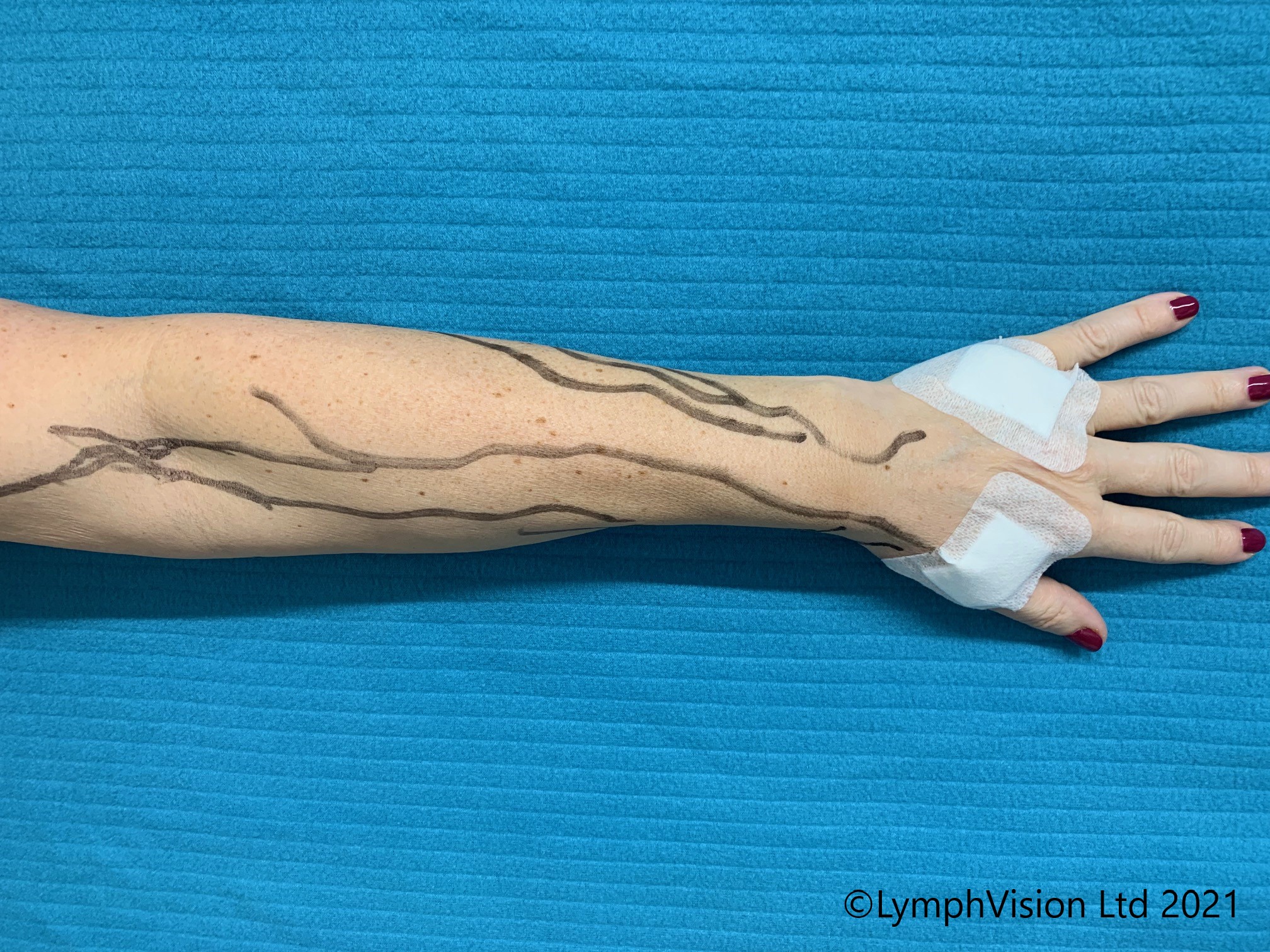
Indocyanine green (ICG) is a water-soluble, near-infrared fluorescent tricarbocyanine dye utilized heavily in clinical medicine, surgical navigation, and diagnostic imaging to visualize blood flow, lymphatic drainage, and organ perfusion in real time.
Introduction to Indocyanine Green (ICG)
Modern medicine relies increasingly on visualization tools that go beyond the naked human eye or standard white-light laparoscopy. Among these, indocyanine green (ICG) has emerged as an indispensable pharmacological agent. Originally approved by the United States Food and Drug Administration (FDA) in the late 1950s for cardiac output and hepatic function testing, ICG has experienced a profound renaissance. Today, it serves as a cornerstone for near-infrared fluorescence (NIRF) imaging in disciplines ranging from oncologic surgery to plastic reconstruction and ophthalmology.
Understanding what ICG is, how it interacts with biological systems, and why it matters requires looking at its unique photophysical properties. When injected into the bloodstream, ICG binds almost exclusively to plasma proteins, confining it primarily to the intravascular space. When illuminated with near-infrared light—typically wavelengths between 750 and 800 nanometers—the dye fluoresces, emitting light at a slightly longer wavelength (around 820 to 850 nanometers).
This fluorescence penetrates tissue to a depth of several millimeters, allowing surgeons and diagnosticians to observe microvascular networks, identify sentinel lymph nodes, and verify tissue viability on live video displays. As healthcare shifts toward minimally invasive and precision procedures, ICG bridges the gap between static preoperative imaging and dynamic, real-time intraoperative guidance.
Core Chemical and Photophysical Properties
The utility of ICG stems directly from its chemical structure and behavior within the human body. As a tricarbocyanine dye, ICG possesses specific absorption and emission spectra that make it uniquely suited for biomedical applications.
| Property | Characteristic |
|---|---|
| Molecular Weight | ~775 g/mol |
| Peak Absorption (Blood) | ~800–805 nm (Near-Infrared Spectrum) |
| Peak Emission | ~825–835 nm |
| Plasma Protein Binding | 95%–98% (primarily albumin and lipoproteins) |
| Elimination Half-Life | 3 to 4 minutes |
| Primary Excretion Route | Biliary / Hepatic system |
Protein Binding and Intravascular Confinement
Upon intravenous administration, ICG molecules rapidly bind to plasma proteins, particularly albumin and alpha-1 lipoproteins. This high degree of protein binding prevents the dye from immediately diffusing through capillary walls into the interstitial space. Consequently, ICG remains within the vascular lumen for a brief period, making it an exceptional tracer for angiography and blood flow assessment.
Near-Infrared Fluorescence Emission
Human tissue contains natural chromophores, such as hemoglobin and water, which absorb visible light heavily and cause significant autofluorescence. However, tissue absorption and scattering are minimized in the near-infrared (NIR) spectrum, particularly in the 700 to 900 nanometer window. By absorbing light at ~805 nm and emitting fluorescence at ~835 nm, ICG operates within this optical window. Specialized laparoscopic and robotic camera systems can capture this emitted NIR light and overlay it as a bright green signal onto the standard white-light surgical field.
Rapid Hepatic Clearance
One of ICG's greatest clinical safety advantages is its pharmacokinetic profile. The liver extracts ICG almost exclusively from the circulation, secreting it unchanged into the bile without undergoing significant intrahepatic metabolism. With a plasma half-life of only 3 to 4 minutes, the dye is cleared rapidly from the body. This rapid elimination allows clinicians to administer multiple doses during a single procedure if successive imaging phases are required, minimizing the risk of cumulative systemic toxicity.
Primary Clinical Applications of ICG
Because ICG highlights blood vessels, lymphatic pathways, and tissue perfusion, its clinical footprint spans several medical specialties. Each application leverages a specific physiological property of the dye.
1. Surgical Oncology and Sentinel Lymph Node Mapping
In cancer surgery, determining whether a tumor has metastasized to regional lymph nodes is vital for staging and treatment planning. ICG is frequently injected around tumor sites to map lymphatic drainage. The dye travels through lymph channels and concentrates in the sentinel lymph nodes—the first nodes receiving drainage from the primary tumor. Surgeons using near-infrared fluorescence can directly visualize these nodes through overlying fatty tissue, reducing the need for radioactive tracers and blue dyes while improving lymph node harvest yields.
2. Hepatobiliary and Liver Surgery
Given that ICG is cleared exclusively by the liver, hepatobiliary surgeons use it in two distinct ways:
- Quantitative Liver Function Assessment: Preoperative clearance tests measure the retention rate of ICG at 15 minutes (ICG-R15). A high retention rate indicates poor hepatic functional reserve, helping surgeons determine if a patient can safely tolerate a major liver resection.
- Negative Staging and Tumor Identification: In certain primary or metastatic liver cancers, tumors lack the normal enzymatic machinery to excrete ICG. When administered intravenously prior to resection, the surrounding healthy liver parenchyma absorbs and excretes the dye, while the tumor retains it or remains unstained, creating a sharp boundary that guides precise anatomical resections.
3. Plastic, Reconstructive, and Vascular Surgery
Flap reconstruction—transferring tissue from one part of the body to another—relies entirely on adequate blood supply. In procedures like breast reconstruction using deep inferior epigastric perforator (DIEP) flaps, surgeons inject ICG intraoperatively to evaluate microvascular perfusion. Areas of the tissue flap that glow brightly receive adequate blood flow, while dark or poorly perfused zones indicate ischemia. Identifying these poorly perfused margins in real time allows surgeons to trim the tissue proactively, preventing postoperative necrosis and flap failure.
4. Ophthalmology and Retinal Angiography
Before the widespread adoption of optical coherence tomography, ICG angiography (ICGA) became a standard diagnostic tool in ophthalmology. Because near-infrared light penetrates the retinal pigment epithelium and choroid better than fluorescein, ICGA allows ophthalmologists to visualize the choroidal vasculature beneath the retina. This is particularly valuable for diagnosing conditions such as choroidal neovascularization, age-related macular degeneration, and inflammatory chorioretinal diseases.
Safety Profile, Contraindications, and Adverse Reactions
While ICG has a decades-long safety record, healthcare providers must evaluate patient-specific risk factors before administration.
Allergic Reactions and Iodine Content
ICG powder is synthesized with sodium iodide to improve solubility, and commercial preparations typically contain up to 5% sodium iodide. Consequently, patients with a documented allergy to iodine or iodinated contrast media historically faced restrictions regarding ICG administration.
However, true allergic reactions to ICG are rare, occurring in roughly 0.05% to 0.3% of administrations. Modern consensus guidelines suggest that a history of shellfish allergy or iodinated contrast allergy is a relative rather than an absolute contraindication, though premedication with corticosteroids and antihistamines may be considered for high-risk patients. Severe anaphylactic reactions, though exceedingly rare, require immediate resuscitation protocols.
Renal and Hepatic Considerations
Unlike iodinated computed tomography (CT) contrast agents, ICG does not cause nephrotoxicity because it is not excreted by the kidneys. Patients with chronic kidney disease can safely receive ICG. Conversely, patients with severe obstructive jaundice or advanced hepatic failure experience delayed clearance, meaning baseline liver function tests should be reviewed prior to diagnostic testing.
Pregnancy and Lactation
Data regarding the use of ICG during pregnancy are limited. It is classified under FDA pregnancy categories as a drug to be used only if clearly needed. Similarly, it remains unknown whether ICG is excreted in human milk; caution is advised when administering ICG to nursing mothers, and temporary cessation of breastfeeding for several hours following administration is sometimes recommended.
Technical Equipment and Intraoperative Integration
The clinical value of ICG is inextricably linked to the hardware used to excite and detect it. A dye injection alone is ineffective without specialized imaging infrastructure.
- Light Sources: Modern surgical light towers incorporate near-infrared laser diodes or filtered xenon/LED arrays that emit light specifically within the 750–800 nm excitation band.
- Sensors and Filters: High-definition laparoscopic cameras and robotic surgical consoles (such as the da Vinci surgical system) utilize specialized optical filters that block visible white light while capturing the faint near-infrared emission signal.
- Display Modes: Surgeons can toggle between standard white-light vision, pure near-infrared monochrome display, and fused color-overlay modes where the fluorescent green signal is digitally merged onto the anatomical field in real time.
The Future of ICG in Precision Medicine
The ongoing evolution of ICG applications centers on molecular targeting and quantitative perfusion analysis. While conventional ICG is a non-targeted fluorophore, researchers are developing bioconjugated ICG derivatives that attach to specific cancer cell surface markers or enzymatic receptors. This targeted fluorescence could allow surgeons to illuminate microscopic tumor margins that are invisible under standard white light or current NIR imaging techniques. Furthermore, software algorithms are being integrated into surgical displays to measure the exact velocity and intensity of ICG uptake, moving subjective visual assessments toward objective, data-driven surgical metrics.
Frequently Asked Questions
What is ICG made of?
ICG is a tricarbocyanine dye officially known as indocyanine green. It is a synthetic, water-soluble compound synthesized with sodium iodide to enhance stability and solubility in aqueous solutions.
Is ICG safe for patients with kidney disease?
Yes. Unlike traditional CT contrast agents that are cleared through the renal system and carry risks of contrast-induced nephropathy, ICG is processed entirely by the liver and excreted through the biliary tract, making it safe for patients with compromised kidney function.
How long does ICG stay in the body?
ICG has a very short plasma half-life of approximately 3 to 4 minutes. It is rapidly bound to plasma proteins, extracted by the liver, and cleared completely from the bloodstream within 15 to 20 minutes.
Does ICG require special surgical equipment?
Yes. Standard surgical lights and standard cameras cannot detect near-infrared fluorescence. To use ICG effectively, operating rooms must be equipped with specialized NIR-capable laparoscopic towers, open surgical scopes, or robotic imaging systems that provide the correct excitation wavelength and capture filters.
Can ICG cause allergic reactions?
While ICG is generally well-tolerated, adverse reactions can occur. Because commercial preparations contain sodium iodide, individuals with known iodine sensitivities should be evaluated carefully. Severe anaphylactic reactions are extremely rare, occurring in less than 0.3% of cases.
Conclusion
Indocyanine green has evolved from a basic cardiovascular and hepatic test into an essential agent for modern surgical and diagnostic navigation. By combining rapid hepatic clearance, high plasma protein binding, and unique near-infrared fluorescence properties, ICG allows clinicians to visualize hidden anatomical structures, assess tissue perfusion, and execute complex procedures with precision. As optical technology and molecular targeting continue to advance, ICG will remain a vital instrument in the ongoing shift toward fluorescence-guided medicine.
Image Gallery
Select an image to open it in a new tab.
Ingrid Bjornson
Global Cyber Policy and Regulation Analyst
