Gram-positive morphology refers to the structural characteristics of bacteria that possess a thick peptidoglycan layer in their cell walls, which retains crystal violet dye during the Gram staining process. These organisms typically appear purple or blue under microscopic examination and are categorized by distinct shapes, such as cocci or bacilli, and specific spatial arrangements.
The Foundations of Gram-Positive Structure
To understand Gram-positive morphology, one must first examine the architecture of the bacterial cell envelope. In microbiology, the distinction between Gram-positive and Gram-negative organisms is not merely a matter of color under a microscope; it reflects fundamental differences in evolutionary biology, metabolic requirements, and pathogenic mechanisms.
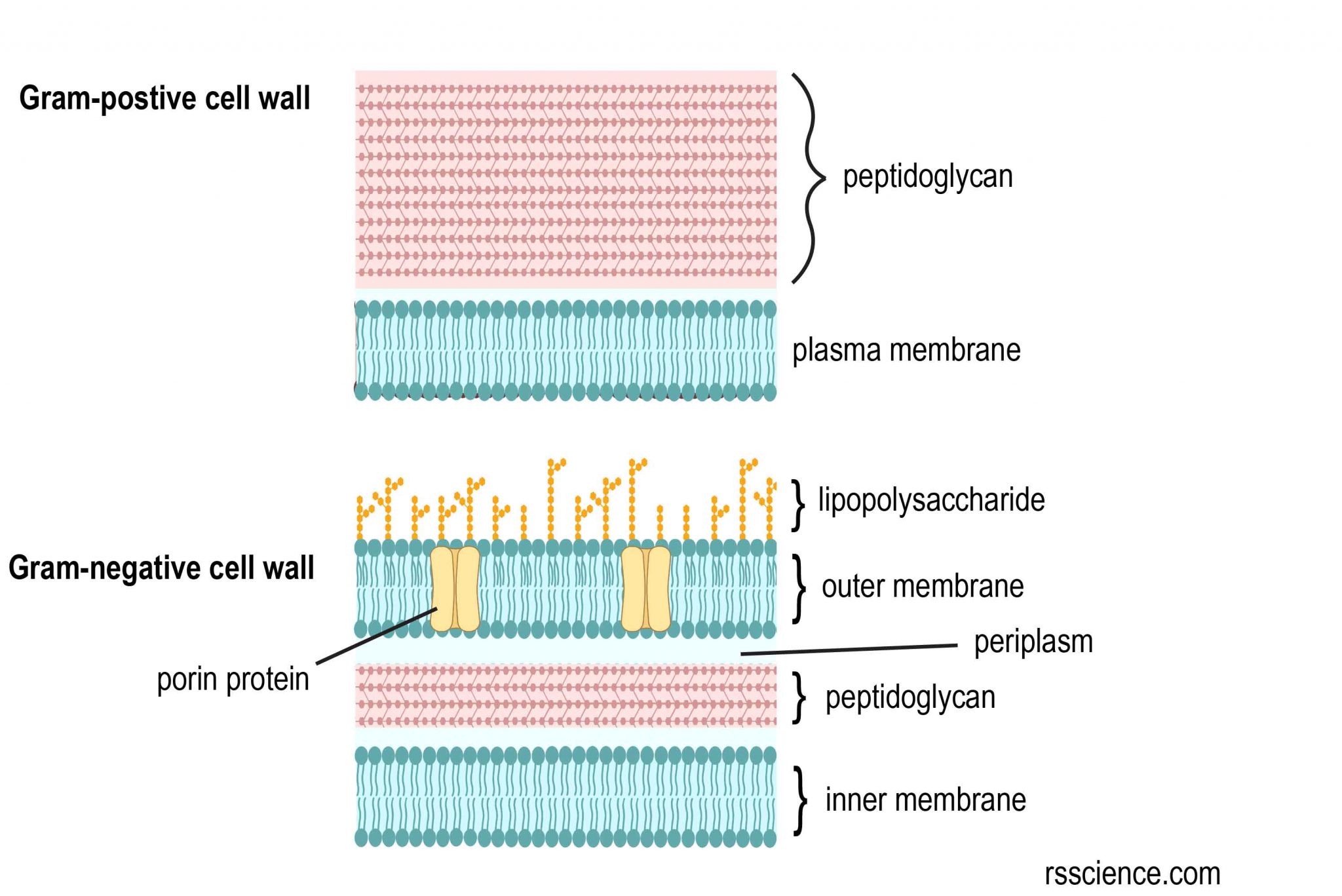
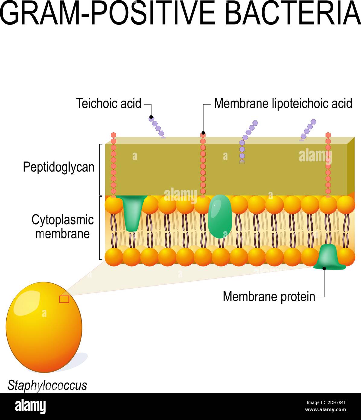
The defining feature of a Gram-positive bacterium is its thick, multilayered cell wall composed primarily of peptidoglycan (also known as murein). This polymer consists of alternating sugars—N-acetylglucosamine (NAG) and N-acetylmuramic acid (NAM)—cross-linked by short peptide chains. In Gram-positive organisms, this peptidoglycan layer can be 20 to 80 nanometers thick, accounting for up to 90% of the cell wall's dry weight.
Beyond peptidoglycan, Gram-positive cell walls contain acidic polysaccharides called teichoic acids. These are either ribitol or glycerol polymers linked by phosphodiester bonds. They are covalently bound to the peptidoglycan (wall teichoic acids) or anchored to the cytoplasmic membrane (lipoteichoic acids). Teichoic acids carry a negative charge, which helps regulate the movement of cations into and out of the cell, provides antigenic specificity, and plays a crucial role in cell wall maintenance and division. Unlike Gram-negative bacteria, Gram-positive bacteria lack an outer lipid membrane and a periplasmic space, making their internal structures more directly exposed to the external environment, yet physically shielded by the dense peptidoglycan mesh.
The Mechanism of Morphological Identification: Gram Staining
The morphological classification of these bacteria relies on the Gram stain, a differential staining technique developed by Hans Christian Gram in 1884. As of 2026, this remains the gold standard for rapid diagnostic microbiology. The procedure involves four primary steps that exploit the thickness of the Gram-positive cell wall.
- Primary Stain (Crystal Violet): All cells are initially stained purple. The dye molecules enter the thick peptidoglycan layer.
- Mordant (Iodine): Iodine is added to form a crystal violet-iodine (CV-I) complex. This complex is larger than the original dye molecule and becomes trapped within the cell wall.
- Decolorization (Alcohol or Acetone): This is the most critical step. In Gram-positive bacteria, the alcohol dehydrates the thick peptidoglycan layer, causing it to shrink and trap the CV-I complexes inside. In contrast, the thin peptidoglycan of Gram-negative bacteria cannot hold the dye, and the outer membrane is dissolved, allowing the dye to wash away.
- Counterstain (Safranin): Since Gram-positive cells are already saturated with purple dye, the red counterstain has no visible effect on them, leaving them purple.
Accurate identification of morphology requires a fresh culture (usually 18–24 hours old). Older cultures may undergo autolysis, where the cell wall begins to break down, leading to "Gram-variable" results where Gram-positive cells incorrectly appear pink (Gram-negative).

Major Morphological Categories: Cocci and Bacilli
Microbiologists categorize Gram-positive bacteria primarily by their shape (morphology) and how they group together (arrangement). These physical traits are genetically determined and serve as the first branch in the taxonomic tree during laboratory identification.
Gram-Positive Cocci
Cocci are spherical or ovoid bacteria. Their arrangement depends on the planes of division during binary fission:
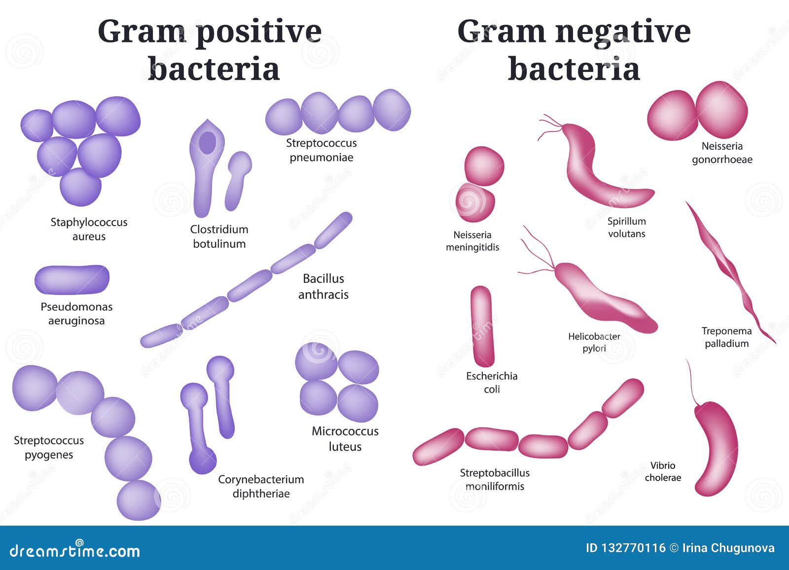
- Clusters (Staphylococci): These divide in multiple planes, resulting in grape-like clusters. Staphylococcus aureus is the most prominent example, often appearing as dense, irregular bunches.
- Chains (Streptococci): These divide in a single plane and remain attached, forming long strings. Streptococcus pyogenes (the cause of strep throat) typically displays this morphology.
- Pairs (Diplococci): Some species, such as Streptococcus pneumoniae, primarily exist in pairs. These are often described as "lancet-shaped" because they are slightly pointed at the ends.
- Tetrads and Sarcina: Some organisms divide in two or three perpendicular planes, forming groups of four (tetrads) or cubical packets of eight (sarcina), such as Micrococcus luteus.
Gram-Positive Bacilli
Bacilli are rod-shaped organisms. Their morphology can vary significantly in length, width, and end-shape (blunt, rounded, or tapered):
- Large, Square-Ended Rods: Typical of the genus Bacillus. When viewed in chains, they may resemble a "bamboo pole" appearance.
- Club-Shaped (Pleomorphic): Some bacilli, such as Corynebacterium diphtheriae, exhibit variations in shape. They may be wider at one end, resembling a club, often arranging themselves in "V" or "L" shapes known as palisades or Chinese letter formations.
- Coccobacilli: These are very short rods that can be easily mistaken for cocci. Listeria monocytogenes is a classic example of a small Gram-positive rod that may appear coccobacillary.
Special Morphological Features: Endospores and Branching
Some Gram-positive bacteria possess specialized morphological features that allow them to survive extreme conditions or mimic other types of organisms like fungi.
Endospore Formation
Two major genera of Gram-positive bacilli, Bacillus and Clostridium, have the ability to form endospores. An endospore is a highly resistant, dormant structure formed within the bacterial cell. Morphologically, the position of the spore can be diagnostic:
- Terminal: At the very end of the rod (e.g., Clostridium tetani, giving it a "drumstick" appearance).
- Subterminal: Near the end but not at the tip.
- Central: In the middle of the cell.
Under a standard Gram stain, spores appear as clear, unstained oval areas within the purple vegetative cell because the thick spore coat resists the dye.
Filamentous and Branching Morphology
Certain Gram-positive bacteria, specifically the Actinomycetes (such as Actinomyces and Nocardia), exhibit a fungus-like morphology. They form long, branching filaments called hyphae. This complex structure is a significant departure from simple rods or spheres and can lead to misidentification if the observer is not familiar with branching Gram-positive morphology.
Comparative Overview of Gram-Positive Morphologies
The following table summarizes the primary morphological characteristics of common Gram-positive genera encountered in clinical and environmental microbiology in 2026.
| Genus | Shape | Arrangement | Distinctive Feature | Clinical Significance |
|---|---|---|---|---|
| Staphylococcus | Cocci | Clusters | Catalase positive | Skin infections, MRSA |
| Streptococcus | Cocci | Chains/Pairs | Catalase negative | Pneumonia, Strep throat |
| Enterococcus | Cocci | Pairs/Short chains | Growth in 6.5% NaCl | UTIs, VRE |
| Bacillus | Bacilli | Chains/Singles | Aerobic spore-formers | Anthrax, Food poisoning |
| Clostridium | Bacilli | Singles/Pairs | Anaerobic spore-formers | Tetanus, C. diff |
| Corynebacterium | Bacilli | Palisades/V-shapes | Pleomorphic "Chinese letters" | Diphtheria |
| Listeria | Coccobacilli | Singles/Short chains | Tumbling motility | Foodborne illness |
| Actinomyces | Filamentous | Branching | Fungus-like filaments | Actinomycosis |
Clinical and Diagnostic Implications of Morphology
In clinical settings, the rapid assessment of Gram-positive morphology is often the first step in directing life-saving therapy. Because the cell wall of Gram-positive bacteria is so different from Gram-negatives, they respond differently to antibiotics.
Antibiotic Sensitivity: Most Gram-positive bacteria are naturally susceptible to beta-lactam antibiotics (like Penicillin) because these drugs specifically target the peptidoglycan synthesis process. However, the emergence of resistant strains like Methicillin-resistant Staphylococcus aureus (MRSA) has complicated this. Understanding the morphology helps the clinician suspect certain organisms—for example, Gram-positive cocci in clusters in a blood culture strongly suggest Staphylococcus, which may require vancomycin if resistance is suspected.
Pathogenesis: The morphology also dictates how the bacteria interact with the host. Encapsulated Gram-positive cocci, like S. pneumoniae, use their polysaccharide capsules to evade phagocytosis by white blood cells. The ability of Bacillus and Clostridium to form spores allows them to persist in hospital environments or soil for years, making them difficult to eradicate through standard disinfection.
Common Mistakes in Morphological Interpretation
Even for experienced microbiologists, several factors can lead to the misinterpretation of Gram-positive morphology:
- Over-decolorization: If the alcohol/acetone is left on the slide for too long, the crystal violet will be washed out even from the thick peptidoglycan layers, making Gram-positive cells appear Gram-negative.
- Smear Thickness: If the bacterial smear is too thick, the decolorizer cannot penetrate effectively, and Gram-negative cells may appear purple (false positive).
- Antibiotic Treatment: Patients already on antibiotics may have bacteria with "damaged" cell walls. These cells may not retain dye properly, leading to bizarre shapes (pleomorphism) or inconsistent staining.
- Heat Fixing: Overheating the slide during the fixing stage can rupture cell walls, distorting the natural shape and arrangement of the bacteria.
Frequently Asked Questions
Why do Gram-positive bacteria turn purple?
Gram-positive bacteria turn purple because their thick peptidoglycan cell wall traps the crystal violet-iodine complex. When the decolorizer (alcohol) is applied, it dehydrates this thick layer, closing the pores and preventing the large dye complexes from escaping. Consequently, the primary purple stain remains visible even after the red counterstain is applied.
Can a Gram-positive bacterium lose its Gram-positive status?
Yes. If a culture is old (senescent), the enzymes within the bacteria (autolysins) begin to break down the peptidoglycan. A weakened cell wall cannot effectively trap the crystal violet-iodine complex, causing the bacteria to appear pink or "Gram-variable." Additionally, certain bacteria called L-forms can lose their cell walls entirely, rendering them unable to be classified by Gram staining.
What is the difference between Staphylococci and Streptococci morphology?
The primary difference lies in their plane of division. Staphylococci divide in multiple planes, resulting in irregular, grape-like clusters. Streptococci divide in a single plane, resulting in beads-on-a-string or linear chain formations. This distinction is a fundamental diagnostic marker in clinical microbiology.
Are all rod-shaped Gram-positive bacteria spore-formers?
No. While many prominent Gram-positive rods like Bacillus and Clostridium are spore-formers, many others are not. For example, Listeria, Corynebacterium, and Lactobacillus are all Gram-positive bacilli that do not produce spores. The presence or absence of spores is a key physiological trait used for further differentiation.
How does cell wall thickness affect antibiotic treatment?
The thick peptidoglycan layer of Gram-positive bacteria is the direct target for many antibiotics, such as penicillins and cephalosporins. These drugs inhibit the cross-linking of the peptidoglycan, causing the cell to burst due to osmotic pressure. Gram-negative bacteria have an outer membrane that acts as a barrier to some of these drugs, which is why Gram-positive morphology often suggests a different range of effective treatments.
Practical Recommendations for Laboratory Observation
To ensure accurate identification of Gram-positive morphology, practitioners should adhere to standardized protocols. Always use cultures that are in the exponential growth phase (typically 18 hours). When performing the Gram stain, ensure the smear is a thin, single layer of cells to allow for even exposure to reagents.
Furthermore, always include a known Gram-positive control (such as Staphylococcus aureus) and a known Gram-negative control (such as Escherichia coli) on the same slide or a parallel slide to verify that the staining reagents and timing are correct. If you observe branching or unusually long filaments, consider extending the incubation time or using specialized media to check for Actinomycetes. In cases where the Gram stain is ambiguous, secondary tests like the Catalase test (to distinguish Staphylococcus from Streptococcus) or the KOH string test can provide additional confirmation of the cell wall type.
Image Gallery
Select an image to open it in a new tab.




Simon Caldwell
Enterprise Technology and Systems Editor
