Understanding the Ligament Wrist Injury: A Complete Guide to Diagnosis and Recovery
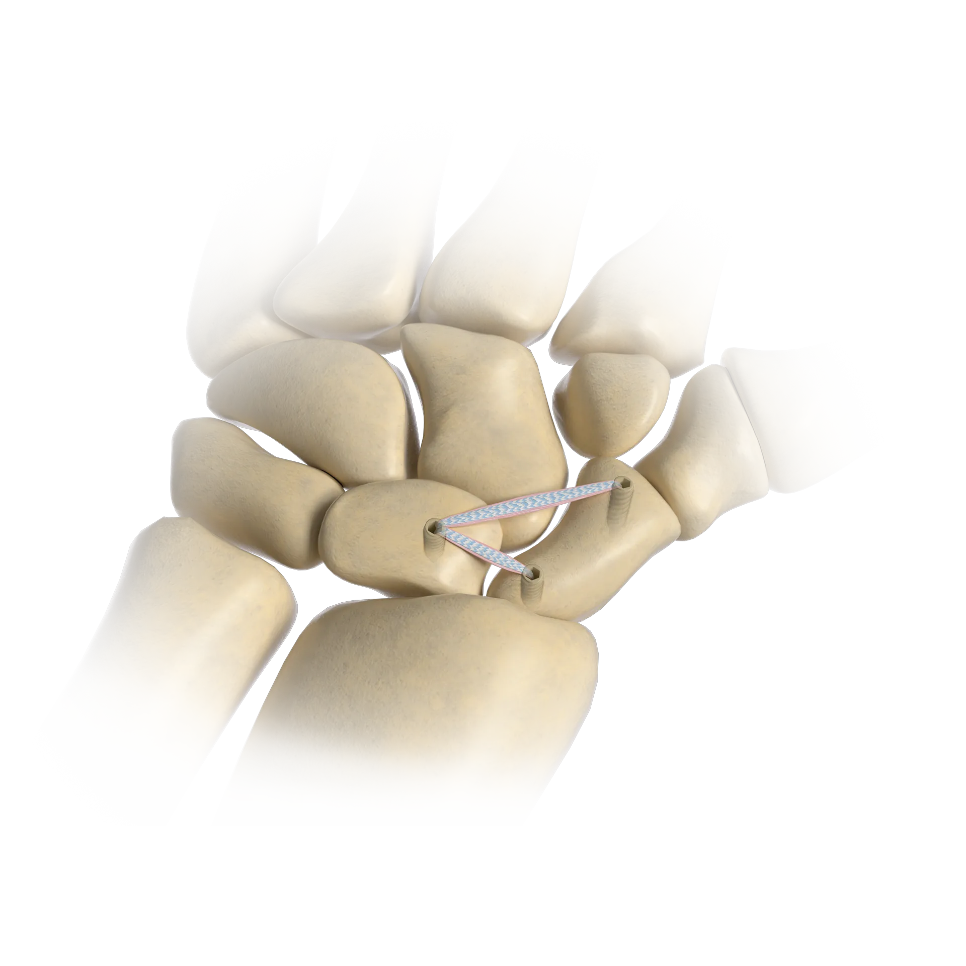
A wrist ligament injury occurs when the tough bands of connective tissue that link the eight carpal bones are stretched or torn, often due to a fall or sudden impact. These injuries range from minor sprains to complete ruptures, frequently requiring clinical evaluation, specialized imaging, and tailored rehabilitation to restore joint stability and prevent long-term arthritis.
The human wrist is one of the most complex joints in the body, consisting of eight small carpal bones arranged in two rows, connected by an intricate network of ligaments. These ligaments are responsible for maintaining the structural integrity of the joint while allowing for the wide range of motion necessary for daily tasks. When one or more of these ligaments are compromised, the entire mechanics of the hand and forearm can be disrupted. As of 2026, medical understanding of wrist biomechanics has led to highly specialized treatment protocols that prioritize early intervention to avoid chronic instability.
The Anatomy of Wrist Ligaments
To understand a wrist ligament injury, one must first appreciate the complexity of the wrist's architecture. Unlike the hinge joint of the knee, the wrist is a "condyloid" joint that allows for flexion, extension, abduction, and adduction. This mobility is facilitated by two primary types of ligaments: intrinsic and extrinsic.
Intrinsic ligaments are located entirely within the carpal bones themselves. These are short, incredibly strong fibers that bind the carpal bones together. The most clinically significant of these is the Scapholunate (SL) ligament, which connects the scaphoid and lunate bones. This ligament is often referred to as the "ACL of the wrist" because it is the primary stabilizer of the proximal carpal row. A tear here can lead to significant instability and a condition known as Scapholunate Advanced Collapse (SLAC).
Extrinsic ligaments connect the carpal bones to the radius and ulna (the forearm bones) or to the metacarpals (the hand bones). These ligaments guide the overall movement of the wrist and provide secondary stability. Another critical structure often grouped with ligamentous injuries is the Triangular Fibrocartilage Complex (TFCC). While technically a fibrocartilage structure, the TFCC functions similarly to a ligament by stabilizing the distal radioulnar joint (DRUJ) on the pinky side of the wrist.
Common Mechanisms of Injury
Most wrist ligament injuries are the result of acute trauma. The most frequent cause is a Fall On an Outstretched Hand (FOOSH). When a person slips, the natural reflex is to put the hand out to break the fall. If the wrist is extended back (hyperextended) upon impact, the force can exceed the tensile strength of the ligaments, leading to a tear.
Beyond falls, other common causes in 2026 include:
- Athletic Injuries: High-impact sports such as football, snowboarding, and gymnastics place extreme stress on the wrist.
- Repetitive Microtrauma: While less common than acute tears, repetitive heavy loading or vibration (common in certain manual trades) can gradually weaken the ligamentous structures.
- Direct Impact: A direct blow to the wrist, often seen in motor vehicle accidents or contact sports, can force the carpal bones out of alignment, rupturing the connecting tissues.
Classification and Grading of Wrist Sprains
Medical professionals categorize ligament injuries into three distinct grades. This grading system is essential for determining the appropriate course of treatment and predicting the recovery timeline.
Grade I: Mild Sprain
In a Grade I injury, the ligament fibers are stretched but not actually torn. The joint remains stable, and there is no significant misalignment of the carpal bones. Patients typically experience localized pain and slight swelling, but they maintain most of their grip strength.
Grade II: Moderate Sprain (Partial Tear)
A Grade II injury involves a partial tear of the ligament fibers. This is a more significant injury that results in some joint laxity (looseness). Pain is more intense, and swelling and bruising are usually present. Functional use of the hand may be limited due to pain and a feeling of instability.
Grade III: Severe Sprain (Complete Tear)
A Grade III injury is a full rupture of the ligament. This often results in gross instability of the wrist joint. In many cases, a Grade III tear of the Scapholunate ligament will cause a visible gap between the bones on an X-ray. These injuries often require surgical intervention because the ligament cannot heal across a significant gap on its own.
Identifying Symptoms and Seeking Diagnosis
The symptoms of a wrist ligament injury can often mimic those of a simple muscle strain or even a minor fracture. However, certain "red flags" suggest a more serious ligamentous issue. Persistent pain on the radial (thumb) side or ulnar (pinky) side that does not improve with rest is a primary indicator.
Key symptoms include:
- Swelling and Bruising: Immediate swelling usually follows the trauma. Bruising may appear within 24 to 48 hours.
- Weakness: A significant drop in grip strength or the inability to bear weight through the palm.
- Popping or Clicking: A mechanical sensation of clicking or "clunking" when moving the wrist, which often indicates that the carpal bones are shifting abnormally.
- Tenderness to Touch: Sharp pain when pressure is applied directly over the site of the ligament (e.g., the "anatomical snuffbox").
Diagnostic Procedures
Because ligaments are soft tissues, they do not show up on standard X-rays. Therefore, the diagnostic process in 2026 involves a tiered approach:
- Physical Examination: A hand specialist will perform provocative tests, such as the Watson Shift Test, to check for instability in the scaphoid bone.
- X-rays: While X-rays don't show the ligament, they show the spacing between bones. A "Terry Thomas sign" (a wide gap between the scaphoid and lunate) is a classic indicator of an SL tear.
- MRI and MR Arthrography: Magnetic Resonance Imaging is the standard for viewing soft tissue. In complex cases, a dye is injected into the joint (arthrography) before the MRI to make small tears more visible.
- Diagnostic Arthroscopy: This is the gold standard. A surgeon inserts a tiny camera into the wrist to directly visualize the ligament and test its tension with a probe.
Treatment Options: From Conservative to Surgical
The choice of treatment depends heavily on the grade of the injury and the specific ligament involved.
Non-Surgical Management
For Grade I and some Grade II injuries, conservative management is highly effective. The primary goal is to protect the ligament while it heals and to reduce inflammation.
- Immobilization: Using a custom-molded splint or a cast for 2 to 6 weeks helps keep the carpal bones in the correct alignment.
- Medication: Non-steroidal anti-inflammatory drugs (NSAIDs) are used to manage pain and swelling.
- Hand Therapy: A certified hand therapist (CHT) guides the patient through range-of-motion exercises and strengthening once the initial healing phase is complete.
Surgical Intervention
If the ligament is completely ruptured (Grade III) or if the joint is unstable, surgery is usually necessary. As of 2026, minimally invasive arthroscopic techniques are preferred whenever possible.
- Ligament Repair: If the injury is acute (less than 6 weeks old), the surgeon may be able to suture the original ligament back together and pin the bones in place with K-wires.
- Ligament Reconstruction: For chronic injuries, the original ligament may be too degraded to repair. In these cases, a tendon graft (often taken from the patient’s own forearm or wrist) is used to reconstruct the ligament.
- Proximal Row Carpectomy or Fusion: In advanced cases where arthritis has already set in, "salvage" procedures like removing the first row of carpal bones or fusing them together may be necessary to eliminate pain, albeit at the cost of some mobility.
Comparison of Common Wrist Ligament Injuries
| Injury Type | Primary Location | Common Symptom | Standard Initial Treatment |
|---|---|---|---|
| Scapholunate (SL) Tear | Thumb side / Central wrist | Pain with weight-bearing; clicking | Splinting or surgical repair |
| TFCC Tear | Pinky side (Ulnar) | Pain with rotation (turning a key) | Bracing, injections, or debridement |
| Lunotriquetral (LT) Tear | Ulnar/Central | Deep aching; weakness | Immobilization or arthroscopic pinning |
| Dorsal Intercarpal Sprain | Top of the wrist | Pain when pushing up from a chair | Rest and NSAIDs |
The Road to Recovery: Rehabilitation Timeline
Recovery from a wrist ligament injury is a marathon, not a sprint. Because ligaments have a relatively poor blood supply compared to muscles, they heal slowly.
Weeks 1–6: Protection Phase The wrist is typically immobilized. For surgical patients, this involves a cast or heavy splint. The focus is on maintaining the mobility of the fingers and shoulder while the wrist remains still.
Weeks 6–12: Controlled Motion Once the initial "knit" of the ligament is established, the splint is removed for controlled exercises. Patients begin "dart-thrower’s motion" (a specific diagonal movement) which puts the least amount of stress on the healing Scapholunate ligament.
Months 3–6: Strengthening Gradual weight-bearing is introduced. Patients work on grip strength and forearm stability. Athletes may begin sport-specific drills but are usually restricted from full-contact activities.
Months 6–12: Full Return to Activity Complete maturation of the ligament tissue can take up to a year. By this point, most patients have regained 80-90% of their original strength and range of motion, though some permanent stiffness is common following major reconstructions.
Long-Term Outlook and Prevention
The most significant complication of an untreated wrist ligament injury is chronic instability. When ligaments fail to hold the carpal bones in their proper tracks, the bones rub together abnormally. This leads to the premature wearing down of cartilage, resulting in osteoarthritis. In the case of SL tears, this follows a predictable pattern known as a SLAC wrist, which eventually requires joint-fusing surgery.
To prevent these injuries, individuals should:
- Use Protective Gear: Wear wrist guards during high-risk activities like skating or snowboarding.
- Improve Ergonomics: Ensure workstations are set up to avoid repetitive strain.
- Maintain Bone Health: Adequate Vitamin D and Calcium intake support the bone density needed to prevent avulsion fractures (where the ligament pulls a piece of bone away).
Professional Advice Limitation
The information provided in this article is for educational purposes only and does not constitute medical advice, diagnosis, or treatment. Wrist injuries can be complex and may involve underlying fractures or nerve damage. Always seek the advice of a qualified healthcare provider or hand specialist regarding any persistent pain or injury. Never disregard professional medical advice or delay in seeking it because of something you have read here.
Frequently Asked Questions
Can a wrist ligament tear heal on its own without surgery?
Minor (Grade I) and some partial (Grade II) tears can heal with proper immobilization and physical therapy. However, a complete (Grade III) rupture of a major stabilizer like the Scapholunate ligament rarely heals on its own because the ends of the ligament are pulled apart by the tension of the surrounding bones. Without surgical intervention to realign the bones, these tears usually lead to chronic instability.
How do I know if I have a sprain or a fracture?
It is often impossible to tell the difference without imaging. Both cause pain, swelling, and loss of function. Generally, a fracture may cause more immediate, intense swelling and visible deformity, whereas a ligament tear might feel more like a deep, unstable ache. Because many wrist fractures (like scaphoid fractures) don't show up on initial X-rays, any significant wrist injury should be evaluated by a specialist.
What is the "Watson Test" used by doctors?
The Watson Test, or Scaphoid Shift Test, is a physical examination technique used to assess the integrity of the Scapholunate ligament. The doctor applies pressure to the scaphoid bone while moving the wrist from side to side. If the ligament is torn, the scaphoid will shift out of its normal position, often causing a "clunk" or pain when the pressure is released.
How long will I be out of work after wrist ligament surgery?
The timeline depends on the physical demands of your job. For sedentary office work, many people return within 1 to 2 weeks, provided they can work with a cast or splint. For manual labor or jobs requiring heavy lifting, the return-to-work timeframe is typically 3 to 6 months, as the ligament must be fully matured to handle significant loads.
Why does my wrist still click months after the injury?
Clicking can be caused by several factors. In some cases, it is simply scar tissue or "crepitus" within the joint. However, persistent, painful clicking often suggests that the carpal bones are not tracking correctly due to ligamentous laxity. If the clicking is accompanied by pain or a feeling of the wrist "giving way," you should consult a hand surgeon for a follow-up evaluation.
Practical Recommendation for Next Actions
If you suspect you have a wrist ligament injury, the immediate priority is immobilization. Use a rigid over-the-counter wrist brace to prevent further movement of the carpal bones. Follow the RICE protocol (Rest, Ice, Compression, Elevation) for the first 48 to 72 hours. If pain persists beyond a few days, or if you experience a loss of grip strength and mechanical clicking, schedule an appointment with an orthopedic hand specialist. Early diagnosis—ideally within the first six weeks—significantly increases the chances of a successful repair rather than a more complex reconstruction.
Image Gallery
Select an image to open it in a new tab.
Omar Farooq
EdTech Innovation and Virtual Pedagogy Journalist
