
An embolism occurs when a moving object in the bloodstream—such as a blood clot, air bubble, or fat globule—becomes lodged in a blood vessel, blocking blood flow. This blockage can cause severe tissue damage or organ failure depending on its location, often requiring immediate medical intervention to prevent life-threatening complications.
Understanding an embolism requires a clear distinction between the blockage itself and the material that causes it. In the medical field, the obstructing object is called an embolus. When this embolus travels through the vascular system and eventually reaches a vessel too narrow to pass through, the resulting state of blockage is known as an embolism. This condition is a leading cause of morbidity and mortality worldwide, particularly in the forms of pulmonary embolisms and ischemic strokes.
The Pathophysiology of an Embolism
To comprehend how an embolism functions, one must first understand the difference between thrombosis and embolism. A thrombus is a blood clot that forms locally within a blood vessel and stays attached to the vessel wall. While a thrombus is dangerous because it can narrow the vessel (thrombosis), it becomes an embolus the moment a piece of it breaks away and enters the general circulation.
The human circulatory system is a complex network of "pipes" that vary in diameter. The heart acts as a pump, pushing blood through large arteries which then branch into smaller arterioles and eventually microscopic capillaries. An embolus typically originates in a larger vessel or the heart itself. As it travels downstream, the vessels inevitably narrow. The embolism occurs at the specific point where the diameter of the embolus exceeds the diameter of the blood vessel.
Once the vessel is blocked, the tissue downstream is deprived of oxygenated blood, a state known as ischemia. If the blockage is not resolved quickly, the deprived tissue begins to die, leading to infarction. This is the biological mechanism behind many "attacks," including myocardial infarctions (heart attacks) and many types of strokes.
Primary Types of Embolism
Embolisms are categorized based on the material that comprises the embolus and the location where the blockage occurs. In 2026, clinical practice continues to prioritize the rapid identification of these specific types to determine the appropriate course of treatment.
Pulmonary Embolism (PE)
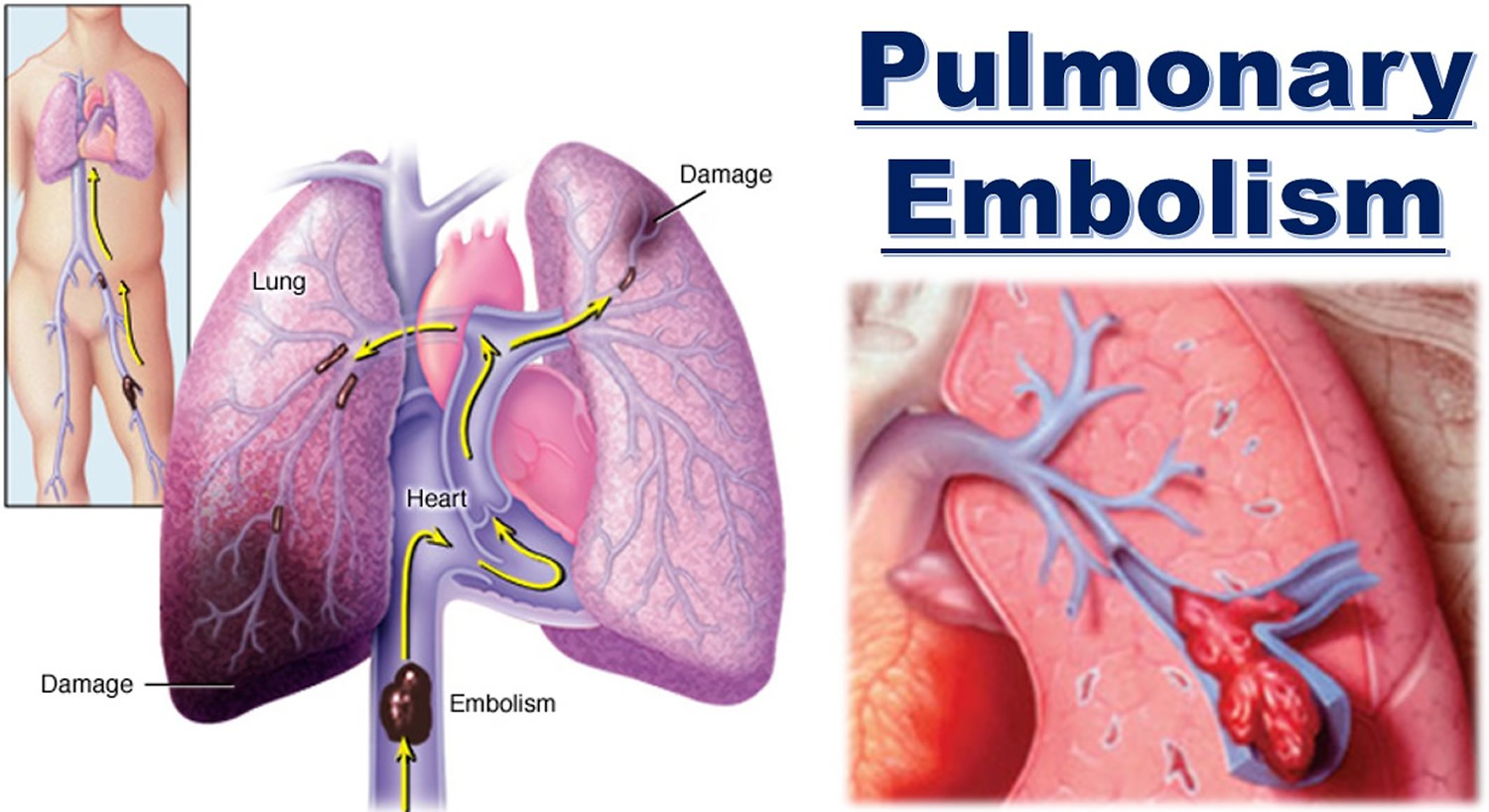
The most common and dangerous form of venous embolism is the pulmonary embolism. This occurs when a clot, usually originating from a Deep Vein Thrombosis (DVT) in the legs, travels through the right side of the heart and lodges in the pulmonary arteries of the lungs. Because the lungs are responsible for oxygenating the entire body’s blood supply, a large PE can lead to sudden heart failure or a dramatic drop in blood oxygen levels.
Arterial Embolism
Unlike pulmonary embolisms, which affect the venous-to-pulmonary circuit, arterial embolisms occur in the systemic arteries. These often originate from the heart (cardioembolism) or from large arteries like the carotid.
- Ischemic Stroke: When an embolus blocks an artery supplying the brain.
- Peripheral Embolism: When an embolus lodges in the extremities, typically the legs, potentially leading to gangrene if untreated.
- Mesenteric Embolism: A blockage in the arteries supplying the intestines, which is a surgical emergency.
Fat Embolism
A fat embolism usually occurs following a traumatic injury, such as a fracture of a long bone (like the femur). When a bone breaks, fat globules from the bone marrow can enter the bloodstream. While many small fat emboli may not cause symptoms, "Fat Embolism Syndrome" can cause respiratory distress, neurological changes, and a characteristic petechial rash (small red spots on the skin).
Air Embolism
An air embolism involves gas bubbles entering the circulatory system. This can happen during surgery, through improperly managed intravenous (IV) lines, or as a result of rapid decompression in scuba diving (the "bends"). The air bubble can act as a physical barrier in the heart or brain, preventing blood flow just as effectively as a solid clot.
Septic Embolism
This occurs when a pocket of infection—containing bacteria and pus—breaks off and travels through the blood. This is most commonly seen in patients with infective endocarditis (infection of the heart valves). These emboli are doubly dangerous because they not only block blood flow but also spread infection to the site of the blockage, often causing abscesses.

Common Causes and Risk Factors
The development of an embolism is rarely a random event; it is usually the result of underlying physiological conditions or lifestyle factors that promote the formation of clots or the entry of foreign material into the blood.
The Virchow’s Triad
Medical professionals use a framework called Virchow’s Triad to understand why blood clots (the most common emboli) form:
- Stasis of Blood Flow: When blood doesn't move, it tends to clot. This is why long flights, bed rest after surgery, or sedentary lifestyles are major risk factors.
- Endothelial Injury: Damage to the lining of the blood vessels (from smoking, high blood pressure, or trauma) creates a "sticky" surface where clots can easily attach.
- Hypercoagulability: Some people have blood that clots more easily than normal due to genetics, cancer, pregnancy, or the use of certain medications like hormonal birth control.
Chronic Conditions
Conditions such as Atrial Fibrillation (AFib) are leading causes of arterial embolisms. In AFib, the upper chambers of the heart quiver instead of beating effectively. This allows blood to pool and clot in the heart. If one of these clots is pumped out, it often heads straight to the brain, causing a stroke. According to the American Heart Association, patients with AFib have a significantly higher risk of stroke than those with a regular heart rhythm.
Recognizing the Symptoms
Symptoms of an embolism vary wildly depending on where the embolus lands. Speed is the most critical factor in survival and recovery.
| Embolism Location | Primary Symptoms | Physical Signs |
|---|---|---|
| Lungs (Pulmonary) | Sudden shortness of breath, sharp chest pain | Rapid heart rate, coughing up blood, low oxygen |
| Brain (Stroke) | Facial drooping, arm weakness, speech difficulty | Confusion, sudden loss of vision, lack of coordination |
| Leg (Peripheral) | Severe pain, numbness | Limb is cold to the touch, pale or blue skin, no pulse |
| Heart (Coronary) | Pressure or squeezing in chest, jaw pain | Sweating, nausea, shortness of breath |
| Intestines (Mesenteric) | Sudden, severe abdominal pain | Nausea, vomiting, "pain out of proportion" to exam |
Diagnostic Procedures and Verification
As of 2026, the gold standard for diagnosing an embolism involves a combination of blood markers and advanced imaging.
- D-Dimer Test: This is a blood test that measures a substance released when a blood clot breaks up. While a high D-dimer doesn't prove an embolism exists, a low D-dimer can often rule one out in low-risk patients.
- CT Angiography (CTPA): For pulmonary embolisms, a CT scan with intravenous contrast is the primary diagnostic tool. It allows doctors to see the actual "filling defects" where the blood cannot flow around the clot.
- Duplex Ultrasonography: This is used to find the "source" of the embolism, specifically checking the deep veins of the legs for existing thrombi.
- Echocardiogram: An ultrasound of the heart can identify clots sitting in the heart chambers or signs of strain on the heart caused by a blockage in the lungs.
Current Treatment Modalities
The management of an embolism focuses on two goals: removing or dissolving the current blockage and preventing new ones from forming.
Anticoagulation Therapy
Most patients with a blood-clot-related embolism are started on anticoagulants, commonly known as blood thinners. In 2026, Direct Oral Anticoagulants (DOACs) like apixaban and rivaroxaban are the standard of care for many. These medications do not actually "thin" the blood or dissolve the existing clot; rather, they prevent the clot from getting larger and stop new clots from forming while the body’s natural processes slowly break the embolus down.
Thrombolytic Therapy ("Clot Busters")
In life-threatening cases, such as a massive pulmonary embolism or an acute ischemic stroke, doctors may use thrombolytics. These are powerful drugs (like tPA or Tenecteplase) delivered via IV that aggressively dissolve clots. However, they carry a high risk of causing internal bleeding and must be used with extreme caution.
Surgical and Interventional Procedures
If medications are insufficient or contraindicated, mechanical intervention is required:
- Embolectomy: A surgeon physically removes the embolus through an incision in the vessel.
- Mechanical Thrombectomy: A catheter-based procedure where a specialist (such as an interventional radiologist) threads a wire through the arteries to "grab" or suction the clot out. This is now a primary treatment for many stroke patients.
- IVC Filters: For patients who cannot take blood thinners, a small metal filter can be placed in the inferior vena cava (the large vein returning blood to the heart) to "catch" clots before they reach the lungs.
Prevention and Lifestyle Adjustments
Preventing an embolism is often more effective than treating one. Most preventative strategies focus on maintaining healthy blood flow and reducing the "stickiness" of the blood.
- Mobility: On long flights or car rides, it is essential to move the legs frequently. Simple "ankle pumps" can keep blood from pooling in the calves.
- Hydration: Dehydration leads to thicker blood, which increases the risk of thrombus formation.
- Compression Wear: For individuals at high risk or those recovering from surgery, graduated compression stockings help the veins return blood to the heart more efficiently.
- Smoking Cessation: Smoking causes significant damage to the endothelium, the internal lining of the blood vessels, making them prone to both plaque buildup and clot attachment.
- Post-Surgical Care: Modern surgical protocols emphasize "early ambulation"—getting patients out of bed and walking as soon as possible after a procedure—to mitigate the high risk of DVT and subsequent PE.
FAQs
How do I know if I have a pulmonary embolism?
Symptoms often appear suddenly and include shortness of breath that feels out of proportion to your activity level, sharp chest pain that worsens when you take a deep breath, and a rapid heart rate. If you also have swelling or pain in one leg (signs of DVT), the likelihood of a pulmonary embolism is higher, and you should seek emergency care immediately.
Can an embolism happen without a blood clot?
Yes. While blood clots are the most frequent cause, embolisms can be caused by air bubbles (from diving or medical errors), fat (from broken bones), cholesterol plaque (from atherosclerosis), or even amniotic fluid during childbirth. These are rarer but can be just as dangerous as a blood-clot embolism.
Is an embolism the same as a stroke?
An embolism is a mechanism, while a stroke is a clinical outcome. Specifically, an embolic stroke is a type of ischemic stroke caused when an embolus travels to the brain and blocks an artery. Not all embolisms are strokes (some land in the lungs or legs), and not all strokes are caused by embolisms (some are caused by local blood vessel ruptures or local clots).
What are the long-term effects of an embolism?
The effects depend on the location and the duration of the blockage. A pulmonary embolism can lead to chronic thromboembolic pulmonary hypertension (high blood pressure in the lung arteries), while a stroke can lead to permanent neurological deficits. However, with prompt treatment and adherence to blood-thinning medications, many people recover fully and go on to live healthy lives.
Why is surgery a risk factor for embolism?
Surgery increases risk for two reasons: physical trauma to blood vessels during the procedure can trigger the body's clotting response, and the subsequent period of immobility (bed rest) allows blood to pool in the legs. Surgeons often prescribe "prophylactic" (preventative) blood thinners or use mechanical leg pumps during and after surgery to counteract these risks.
Summary and Next Actions
If you suspect you or someone else is experiencing an embolism—characterized by sudden shortness of breath, chest pain, or one-sided weakness—call emergency services immediately. Do not attempt to drive yourself to the hospital, as symptoms can worsen rapidly.
For those concerned about their long-term risk, the most effective next action is to consult with a primary care provider to evaluate your cardiovascular health. Discuss your family history of blood clots, manage your blood pressure, and if you are planning a long trip or surgery, ask about specific DVT prevention strategies. Regular physical activity remains the most potent defense against the stasis that leads to most embolic events.
Image Gallery
Select an image to open it in a new tab.




Lars Thomsen
Global Labor Markets and Economic Strategy Editor
