Understanding Glioblastoma Surgery: A Comprehensive Guide to Neurosurgical Intervention
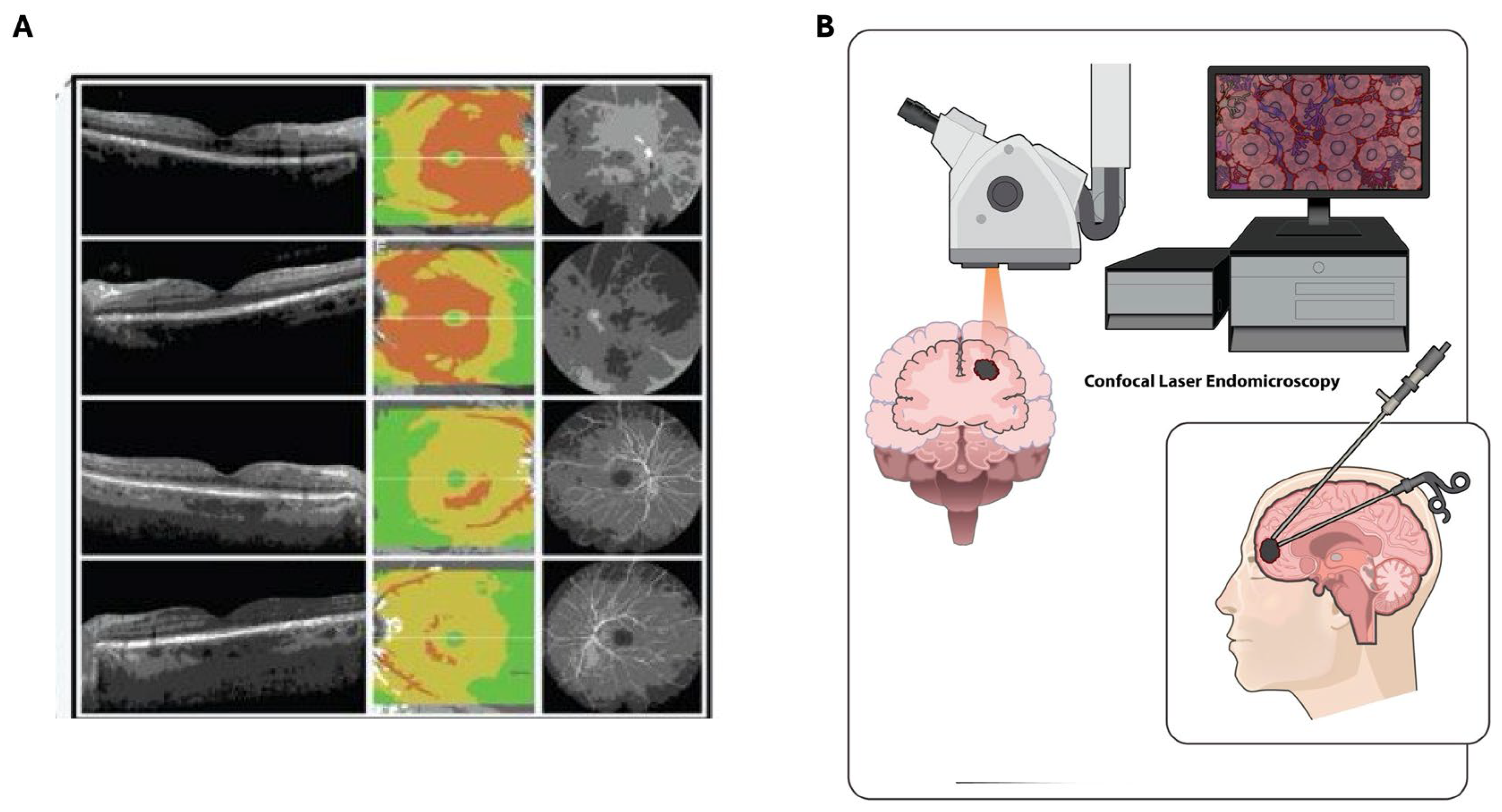
Glioblastoma surgery is a neurosurgical procedure aimed at removing as much of a glioblastoma—a fast-growing, aggressive primary brain tumor—as safely possible. Often serving as the initial step in a multidisciplinary treatment plan, the primary objective of this surgery is to reduce tumor mass, alleviate neurological symptoms, and provide tissue for definitive molecular diagnosis.
The Role of Neurosurgery in Glioblastoma Management
Glioblastoma (GBM) is classified as a Grade 4 astrocytoma, characterized by its rapid cellular proliferation and ability to infiltrate surrounding brain tissue. Because these tumors are diffuse, they lack well-defined borders, making a "cure" via surgery alone mathematically and biologically impossible with current technology. However, surgery remains the cornerstone of standard treatment protocols.
The surgical strategy for glioblastoma is formally referred to as "maximal safe resection." This term is critical to understanding the surgeon's intent: the goal is to remove the greatest possible volume of tumor while preserving the patient’s functional integrity, including speech, motor control, memory, and personality. Sacrificing neurological function to achieve a complete visual resection is generally avoided, as it significantly impairs the patient's quality of life and subsequent ability to tolerate chemotherapy and radiation therapy.
The Clinical Workflow: From Diagnosis to Surgery
Before a patient is scheduled for a craniotomy, a rigorous diagnostic process occurs. This typically involves advanced neuroimaging, most commonly contrast-enhanced Magnetic Resonance Imaging (MRI). In 2026, standard of care includes structural MRI sequences, often supplemented by functional MRI (fMRI) and Diffusion Tensor Imaging (DTI) to map critical eloquent areas of the brain—regions responsible for language, motor function, and sensory processing.
Prerequisites for Surgical Candidates
Not every patient with a brain mass is an immediate candidate for open surgery. Eligibility is determined based on:
- Tumor Location: If the tumor resides in a deep, inaccessible region, such as the thalamus or brainstem, surgery may be deemed too high-risk.
- Performance Status: The patient’s Karnofsky Performance Status (KPS) score is used to assess their ability to function independently and tolerate major surgery.
- Comorbidities: The patient’s cardiac, respiratory, and coagulation health are evaluated to ensure they can survive general anesthesia and the inflammatory response that follows a craniotomy.
Surgical Techniques and Technological Adjuncts
Modern neurosurgery utilizes various technologies to maximize the precision of tumor removal. During a craniotomy, the surgeon gains access to the brain by removing a bone flap. Once the dura (the membrane covering the brain) is opened, the following tools are frequently employed:
- Neuronavigation: Think of this as a "GPS for the brain." Preoperative MRI data is registered to the patient's physical anatomy in the operating room. This allows the surgeon to see exactly where their surgical instruments are in relation to the tumor and critical structures in real-time.
- Intraoperative Fluorescence (5-ALA): 5-Aminolevulinic acid is a metabolic agent administered to patients before surgery. It causes tumor cells to fluoresce (glow) under specific wavelengths of blue light, helping the surgeon distinguish between the tumor and healthy, non-infiltrating brain tissue.
- Intraoperative Neuromonitoring (IONM): During the procedure, neurophysiologists monitor electrical signals from the brain and spinal cord to ensure that the surgeon is not inadvertently damaging pathways essential for movement or speech.
- Awake Craniotomy: For tumors near speech or motor centers, the surgeon may keep the patient awake for parts of the procedure. By engaging the patient in conversation or motor tasks while stimulating the brain, the team can map function in real-time and avoid resecting eloquent cortex.
The Phases of the Procedure
A glioblastoma craniotomy is an intensive, highly orchestrated event involving a large team of specialists.
- Anesthesia Induction: The patient is placed under general anesthesia, ensuring they are immobile and unconscious for the duration of the procedure.
- Positioning and Fixation: The head is secured in a rigid frame (headrest) to ensure stability for the duration of the surgery.
- Craniotomy: The surgeon makes an incision in the scalp and uses a surgical drill to remove a piece of the skull.
- Resection: Using microscopic visualization and neuronavigation, the surgeon identifies the tumor mass and begins the careful process of decompression and resection.
- Closure: Once the tumor is removed, the dura is closed, and the bone flap is replaced and secured with small titanium plates or wires. The scalp is sutured shut.
Expected Outcomes and Recovery
Post-surgery, the patient is typically monitored in an Intensive Care Unit (ICU) for 24 to 48 hours. The most immediate risk is postoperative swelling (edema), which is managed with medications like dexamethasone.
The "success" of the surgery is measured by the extent of resection (EOR). An MRI is typically performed within 48 to 72 hours of the procedure to determine if any residual tumor remains. This baseline image is essential for the oncology team to plan the subsequent phases of care, which generally involve radiation therapy and temozolomide chemotherapy.
| Metric | Purpose |
|---|---|
| Extent of Resection (EOR) | Defines the percentage of the tumor removed; higher EOR is correlated with better survival outcomes. |
| Karnofsky Performance Status (KPS) | Measures the patient's functional ability post-surgery to determine eligibility for clinical trials and therapy. |
| Molecular Profiling | Analysis of the removed tissue for genetic markers (like MGMT methylation or IDH mutation) which dictate prognosis and treatment response. |
Risks and Complications
While neurosurgery has advanced significantly, the brain is a delicate organ, and glioblastoma surgery carries inherent risks. Complications can include:
- Neurological Deficits: New weakness, speech difficulty, or vision loss if the tumor's resection involves eloquent brain tissue.
- Seizures: Even for patients without a prior history of epilepsy, the surgical trauma can trigger seizures, often managed with prophylactic anti-seizure medications.
- Infection: Although rare, surgical site infections or meningitis are potential risks.
- Hematoma: Bleeding in the surgical cavity is an emergency that may require immediate intervention.
Frequently Asked Questions (FAQ)
Does surgery cure glioblastoma? No, surgery is not a cure. Because glioblastoma cells are highly invasive and spread into healthy brain tissue that appears normal on MRI, they cannot be completely excised. Surgery is used to remove the "bulk" of the tumor, which improves symptoms and enhances the effectiveness of other treatments.
How long is the recovery period after surgery? Physical recovery from the craniotomy itself typically takes several weeks. However, the overall recovery process is influenced by the patient’s subsequent treatment plan (radiation and chemotherapy), which usually begins about three to four weeks after the surgery.
Can a tumor recur after surgery? Yes, almost all glioblastomas recur. This is due to the nature of the disease, where single cells left behind after surgery eventually proliferate into a new mass. Follow-up MRIs are performed every two to three months to monitor for recurrence.
Is radiation required after surgery? Yes. Following surgery, the standard of care in 2026 involves a combination of radiation therapy and oral chemotherapy (temozolomide). This combination is designed to target the microscopic tumor cells that could not be seen or removed during surgery.
What is "maximal safe resection" mean for me? It means your surgeon will prioritize your quality of life. If removing a small piece of tumor would likely result in the loss of your ability to walk or speak, the surgeon will typically leave that piece behind to preserve your function, balancing the risks and benefits.
Navigating the Future of Your Care
Following surgery, it is vital to coordinate closely with a neuro-oncology team. This team will handle the interpretation of pathology reports—which categorize the tumor's molecular and genetic profile—and determine the specific chemotherapy and radiation schedule that offers the best outcome for your specific case.
Next steps involve scheduling your first postoperative neuro-oncology appointment, ensuring your medication regimen for seizure prophylaxis and steroids is strictly followed, and arranging for necessary physical or speech therapy support. Maintaining a detailed log of your recovery symptoms and communicating these clearly to your oncology team remains the most effective way to manage your care in the weeks following your procedure.

Image Gallery
Select an image to open it in a new tab.
Omar Farooq
EdTech Innovation and Virtual Pedagogy Journalist
