Why the Sacrum Is Classified as an Irregular Bone: Structure, Function, and Clinical Importance
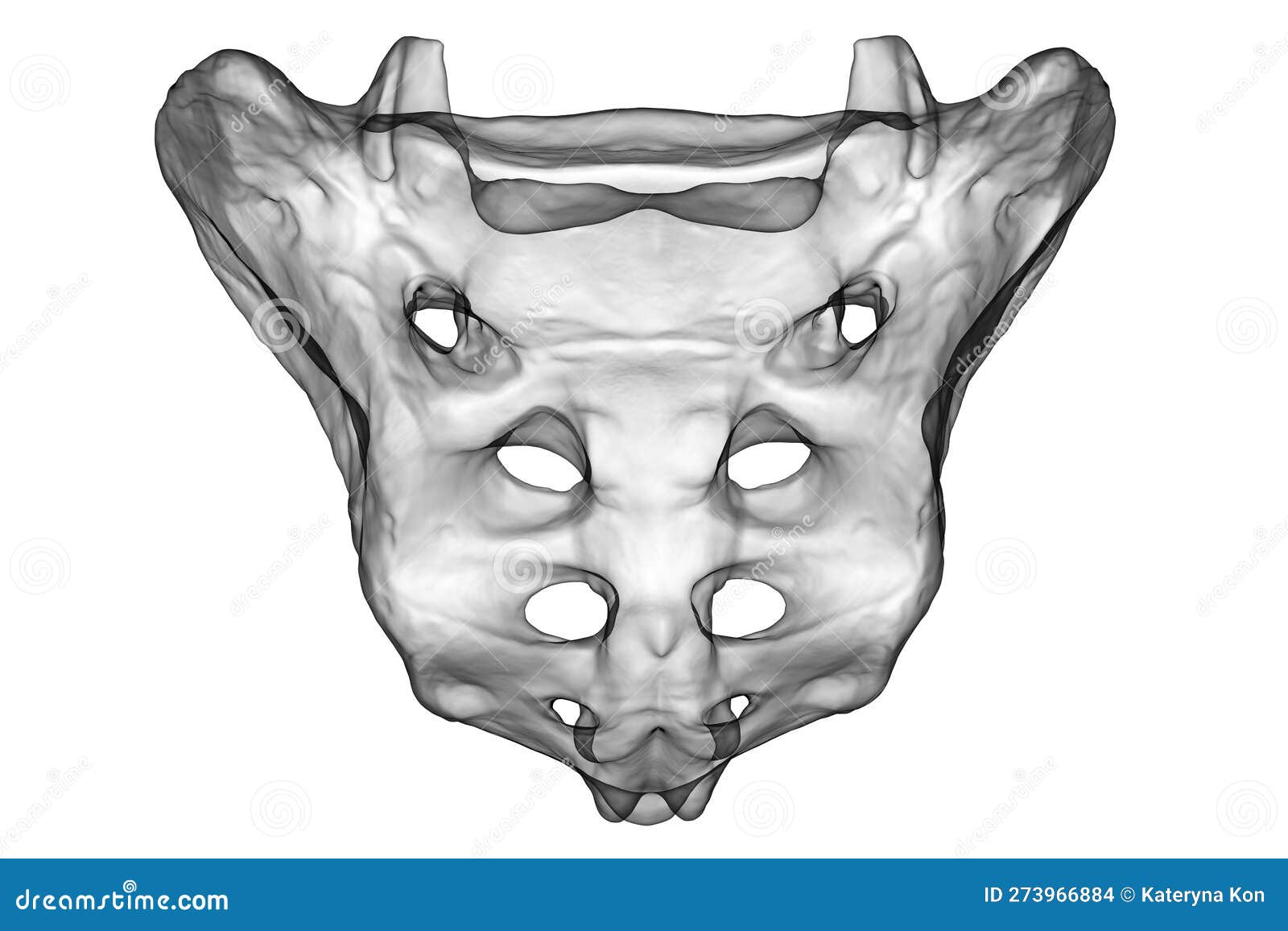
The sacrum is an irregular bone located at the base of the spine, formed by the fusion of five sacral vertebrae (S1–S5). Classified as irregular due to its complex triangular shape, central canal, openings for nerves, and specialized articular surfaces, the sacrum serves as the primary structural bridge connecting the upper axial skeleton to the lower pelvic girdle.
Medical Disclaimer: The anatomical, clinical, and physiological information presented in this article is intended for educational purposes only and does not constitute medical advice. Consult a qualified healthcare professional for clinical diagnoses, injury evaluation, or medical guidance.
Anatomical Classification: Defining the Irregular Bone Category
In human anatomy, the adult skeletal system is categorized into five basic morphological types based on shape and structural characteristics: long bones (such as the femur and humerus), short bones (such as the carpals and tarsals), flat bones (such as the sternum and cranial bones), sesamoid bones (such as the patella), and irregular bones.
Bones are designated as irregular when their complex, non-uniform geometry prevents them from fitting cleanly into any of the other four categories. Rather than possessing a simple shaft, flat expanse, or rounded nodular shape, irregular bones typically feature a combination of specialized structural elements, including short cortical walls, internal spongy bone networks, multiple neural arches, spinous processes, and articular facets.
The sacrum is an irregular bone because it represents a consolidated structure composed of five individually modified vertebral segments. Its architecture integrates a thick central body, lateral expansion wings (alae), posterior neural crests, and multiple neural foramina. This complex geometry is tailored specifically to accommodate spinal nerves, absorb heavy gravitational loads, and provide attachment points for powerful core and hip musculature.
Detailed Anatomical Landmarks of the Sacrum
The sacrum is positioned inferior to the fifth lumbar vertebra (L5) and superior to the coccyx. It forms the posterior wall of the pelvic cavity and participates in the formation of the sacroiliac (SI) joints laterally. Understanding its distinct structural regions demonstrates why its geometry is so intricate.
- L5 Vertebra
- Sacral Promontory (S1 Superior Margin)
- Sacral Ala (Lateral Wings)
- Anterior Sacral Foramina (Pelvic Surface) (S nerve exits)
- Apex of Sacrum
- Coccyx
- Sacral Promontory (S1 Superior Margin)
The Base and Sacral Promontory
The superior aspect of the sacrum, known as the base, articulates with the L5 vertebra. The prominent anterior lip of the S1 vertebral body is termed the sacral promontory. This landmark forms an essential anatomical boundary of the pelvic inlet, which obstetrians measure to assess the dimensions of the birth canal. On either side of the base are the sacral alae (singular: ala), large fan-shaped lateral projections formed by the fusion of the ancestral transverse processes and costal elements of the S1 vertebra.
The Pelvic (Anterior) Surface
The pelvic surface of the sacrum is smooth and concave, facing anteriorly and inferiorly into the pelvic cavity. It is marked by four horizontal transverse lines, which indicate the historical lines of fusion between the five original sacral vertebral bodies. At the lateral ends of these lines lie four pairs of anterior (pelvic) sacral foramina. These smooth openings allow for the passage of the anterior rami of the sacral spinal nerves (S1–S4) and associated blood vessels into the pelvis.
The Dorsal (Posterior) Surface
The dorsal surface of the sacrum is convex, rough, and highly complex, reflecting the attachment of spinal ligaments and deep back muscles such as the erector spinae and multifidus. Key posterior structures include:
- Median Sacral Crest: Formed by the fusion of the spinous processes of the upper sacral vertebrae.
- Intermediate Sacral Crests: Located laterally to the median crest, these are formed by the fusion of the articular processes.
- Lateral Sacral Crests: Formed by the fusion of the transverse processes of the sacral vertebrae.
- Posterior Sacral Foramina: Four pairs of openings lateral to the intermediate crests that allow the posterior rami of the sacral spinal nerves to exit.
- Sacral Canal: The internal continuation of the vertebral canal, housing the nerve roots of the cauda equina (specifically the sacral and coccygeal spinal nerve roots).
- Sacral Hiatus: An inferior U-shaped opening at the terminus of the sacral canal, created by the failure of the S4 and S5 lamina to fuse posteriorly. It is flanked by two prominent projections known as the sacral cornua.
The Lateral Surfaces and Auricular Facets
The lateral margin of the sacrum contains the auricular surface, an ear-shaped cartilage-covered articular facet that joins with the ilium of the pelvis to form the sacroiliac joint. Posterior to the auricular surface lies the sacral tuberosity, a rough, indented region dedicated to the attachment of the powerful interosseous sacroiliac ligaments.

Structural Comparison Across Skeletal Categories
To appreciate why the sacrum is classified as an irregular bone, it is helpful to evaluate its characteristics against the other four primary bone categories.
| Bone Category | Dominant Shape & Geometry | Primary Structural Composition | Typical Biomechanical Role | Representative Examples | Sacrum Comparison |
|---|---|---|---|---|---|
| Long Bones | Tubular, elongated shaft (diaphysis) with expanded ends (epiphyses). | Thick outer compact bone wall surrounding a medullary cavity filled with bone marrow. | Lever arms for locomotion and weight support. | Femur, Tibia, Humerus, Radius | Unlike long bones, the sacrum lacks a single tubular shaft, medullary cavity, or lever mechanics. |
| Short Bones | Cube-like, roughly equal in length, width, and thickness. | Thin layer of compact bone enclosing a interior core of spongy (cancellous) bone. | Shock absorption and multi-directional glide flexibility. | Carpals (wrist), Tarsals (ankle) | Unlike short bones, the sacrum exhibits distinct processes, neural arches, and intricate curved ridges. |
| Flat Bones | Thin, flattened, often gently curved plates. | Two parallel layers of compact bone (tables) sandwiching an inner layer of spongy bone (diploe). | Protection of vital organs and expansive muscular attachment. | Sternum, Ribs, Scapula, Parietal bone | While parts of the sacral ala are somewhat flat, its 3D central canal and neural arches prevent flat bone classification. |
| Irregular Bones | Complex, specialized shapes with variable processes and interior arrangements. | Mixed compact shell with internal cancellous bone optimized for complex stress lines. | Spinal protection, neural transit, weight transfer across axes. | Sacrum, Vertebrae, Sphenoid, Ethmoid, Os Coxae | The sacrum fits here: It combines fused vertebral arches, crests, foramina, and joint facets into a complex whole. |
| Sesamoid Bones | Small, rounded or nodular bones embedded within tendons. | Dense cortical rim with localized spongy tissue. | Altering mechanical pull angle of tendons; reducing friction. | Patella, Pisiform | The sacrum does not develop within a muscle tendon; it is an intrinsic part of the axial skeleton. |
Embryological Development and Fusion Timeline
The sacrum begins its development early in embryonic life as five distinct vertebrae derived from paraxial mesoderm somites. Understanding its developmental path helps clarify how multiple independent segments transform into a single irregular bone.
Developmental Timeline of the Sacrum:
- Embryonic Period: Primary ossification centers form in bodies & arches.
- Birth to Age 5: Neural arches fuse to bodies; lateral components merge.
- Adolescence (16–18): Primary sacral disc spaces begin inter-segmental fusion.
- Young Adulthood (25): Complete consolidation into a single irregular bone.
Primary and Secondary Ossification
Each sacral segment develops through endochondral ossification, where a cartilage model is gradually replaced by bone:
- Primary Ossification Centers: During the third month of fetal development, three primary ossification centers appear for each sacral segment: one for the centrum (vertebral body) and two for the neural arch halves. Additional primary centers develop for the lateral alar components of the upper segments.
- Secondary Ossification Centers: During puberty, secondary ossification centers appear along the outer margins of the sacral bodies, the auricular surfaces, and the lateral borders of the alae.
The Fusion Process
Between ages 16 and 18, the hyaline cartilage discs separating the individual sacral bodies begin to ossify, starting from the inferior segments (S4–S5) and progressing superiorly toward S1. By approximately age 25 to 30, the five segments are fully consolidated into a single solid osseous structure.
Common Developmental Variations
Because sacrum formation relies on the fusion of five separate embryonic units, congenital variations are frequent:
- Sacralization of L5: The fifth lumbar vertebra partially or completely fuses with the S1 segment of the sacrum. This can reduce lumbar spine mobility and alter stress distribution on upper lumbar discs.
- Lumbarization of S1: The S1 segment fails to fuse with S2 and instead remains an independent, mobile vertebra, creating six functional lumbar vertebrae.
- Spina Bifida Occulta: Occurs when the left and right neural arches of S1 or S4–S5 fail to fuse completely along the posterior midline, leaving a small gap in the bone without spinal cord herniation.
Biomechanical Role in Weight Distribution and Pelvic Motion
The sacrum plays a fundamental mechanical role as the keystone of the human pelvis. Positioned between the two hip bones (ossa coxae), it functions as the bridge connecting the vertical axial skeleton to the lower extremities.
- Axial Body Weight (Spine)
- Sacral Base
- Left Ilium (SI Joint Force)
- Acetabulum
- Femoral Heads / Lower Extremities
- Acetabulum
- Right Ilium (SI Joint Force)
- Acetabulum
- Femoral Heads / Lower Extremities
- Acetabulum
- Left Ilium (SI Joint Force)
- Sacral Base
Load Transfer Mechanics
When standing upright, body weight from the head, neck, trunk, and upper limbs travels down through the lumbar spine to rest upon the base of the S1 vertebra. The sacrum transfers this load laterally across its alae and through the sacroiliac joints into the pelvic ring, directing weight to the femoral heads when standing or to the ischial tuberosities when sitting.
The wedge shape of the sacrum contributes to its mechanical stability. Driven downward into the pelvic ring by body weight, its tapered shape naturally wedges it tighter between the iliac bones, a structural mechanism known as form closure.
Sacroiliac Joint Dynamics: Nutation and Counter-Nutation
Although the sacroiliac joint is a amphiarthrodial (slightly movable) joint stabilized by dense interosseous and sacrotuberous ligaments, small planar movements occur around a transverse axis:
- Nutation (Forward Nodding): The sacral base tilts anteriorly and inferiorly while the apex moves posteriorly. Nutation increases pelvic outlet diameter during labor and enhances joint stability during heavy loading (such as landing from a jump).
- Counter-Nutation (Backward Nodding): The sacral base moves posteriorly and superiorly while the apex moves anteriorly. This movement occurs during pelvic recoil and extension of the lower spine.
For further anatomical context on bone classification, consult the comprehensive skeletal system resources available at the National Center for Biotechnology Information (NCBI).
Sexual Dimorphism in Sacral Architecture
The morphology of the sacrum differs noticeably between biological sexes due to evolutionary adaptations for childbirth.
Male Sacrum:
- Narrower
- Longer
- More Curved
Female Sacrum:
- Wider
- Shorter
- Flatter
Width and Proportion: The female sacrum is proportionately wider and shorter than the male sacrum. The S1 articular surface occupies less than half the total width of the sacral base in females, whereas in males, the S1 body occupies more than half the total base width.
Curvature: The male sacrum exhibits a uniform, deep curvature along its entire anterior surface. The female sacrum is relatively flat superiorly and curves more abruptly at its inferior end (S4–S5), creating a roomier pelvic cavity to facilitate childbirth.
Orientation of Auricular Surfaces: The auricular surfaces in females are oriented more obliquely, which allows for greater mobility during late pregnancy under the influence of the hormone relaxin.
Clinical Significance and Pathologies
Because the sacrum contains spinal nerve passages, bears high mechanical loads, and articulates with the pelvic girdle, it is susceptible to structural, metabolic, and traumatic conditions.
Sacroiliac Joint Dysfunction and Sacroiliitis
Inflammation of one or both sacroiliac joints (sacroiliitis) is a hallmark feature of spondyloarthropathies, such as ankylosing spondylitis. Patients experience deep gluteal pain, stiffness, and referred discomfort down the posterior thigh. Mechanical dysfunction can also arise from leg length discrepancies, gait abnormalities, or pregnancy-induced ligamentous laxity.
Sacral Stress Fractures and Traumatic Fractures
Sacral fractures are generally categorized as insufficiency fractures or high-energy traumatic fractures:
- Insufficiency Fractures: Common in elderly individuals with osteoporosis or patients with severe vitamin D deficiency. Minor stress from walking can produce micro-fractures in the sacral alae, causing severe low back or buttock pain.
- Traumatic Fractures: Caused by high-energy impacts such as motor vehicle accidents or falls from heights. Surgeons categorize these using the Denis Classification System:
- Zone 1 (Alar Region): Fractures occur lateral to the sacral foramina. Neurological deficits are rare (~6% of cases).
- Zone 2 (Foraminal Region): Fractures pass through one or more sacral foramina. May cause sciatica or unilateral motor/sensory deficits (S1–S2 nerve roots).
- Zone 3 (Central Canal Region): Fractures extend into the central sacral canal. High risk (~50–60% of cases) of cauda equina injury, causing loss of bowel, bladder, or sexual function.
Caudal Anesthesia and Epidural Injection
The sacral hiatus provides a reliable anatomical access point for regional anesthesia. Clinicians palpate the sacral cornua to locate the hiatus and insert a needle into the lower caudal epidural space. This technique, known as a caudal block, is widely used in pediatric surgery for lower abdominal and lower extremity procedures, as well as in adult pain management for managing chronic radiculopathy.
Frequently Asked Questions
Is the sacrum considered a single bone or five separate bones?
In an adult human, the sacrum is formally classified as a single irregular bone because its constituent vertebrae have fully fused. However, in pediatric and developmental anatomy, it is described as five individual sacral vertebrae (S1–S5) separated by cartilaginous discs.
Why is the sacrum not classified as a flat bone despite its wide appearance?
While the sacral alae present broad, relatively flat lateral surfaces, the overall 3D structure of the sacrum includes an internal spinal canal, irregular bony crests, complex articular processes, and functional nerve foramina. These three-dimensional structural characteristics place it squarely in the irregular bone category.
What is the sacral promontory and why is it clinically important?
The sacral promontory is the prominent anterior projection of the S1 vertebral body. It serves as an essential anatomical landmark for measuring the pelvic inlet dimensions during obstetric evaluations and acts as a primary anchor point during pelvic organ prolapse surgeries such as sacrocolpopexy.
What nerves pass through the sacral foramina?
The anterior sacral foramina transmit the anterior rami of spinal nerves S1 through S4, which form the primary components of the sacral plexus supplying the hip, leg, and pelvic organs. The posterior sacral foramina transmit the posterior rami of the same spinal nerves, which innervate the skin and deep muscles of the dorsal pelvic region.
How does sacral fusion affect spinal mobility?
Because the sacral segments are fused into a rigid unit, there is no inter-vertebral motion between S1 and S5. This rigidity creates a stable platform for weight transfer. Flexibility is concentrated at the lumbosacral junction (L5–S1) above and the sacroiliac joints to a minor degree laterally.
Practical Takeaways and Next Steps
- Recognize Structural Identity: Remember that the sacrum is an irregular bone because its composite structure—formed by five fused vertebrae—combines neural canals, surface openings, and complex joint interfaces that do not fit long, short, flat, or sesamoid categories.
- Understand Load Transfer: The primary biomechanical purpose of the sacrum is to transmit upper body weight laterally into the pelvic girdle via the sacroiliac joints while protecting the sacral spinal nerve roots.
- Evaluate Back and Pelvic Pain: Deep lower back, gluteal, or groin pain should be evaluated with consideration of the sacrum, as conditions ranging from SI joint inflammation to sacral insufficiency fractures can mimic lumbar disc pathology.
- Review Related Concepts: Explore anatomical resources detailing the sacral plexus, pelvic inlet measurements, and lumbar spine mechanics to gain a complete understanding of how the sacrum functions within the broader musculoskeletal system.
Image Gallery
Select an image to open it in a new tab.

Declan O'Connor
Higher Education and Campus Culture Reporter
